- EMDB-40245: Human ER membrane protein complex (EMC) in GDN, 9-subunit map -
+
データを開く
IDまたはキーワード:
読み込み中...
-
基本情報
登録情報
データベース: EMDB / ID: EMD-40245
タイトル
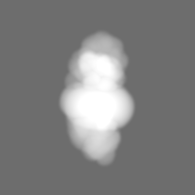
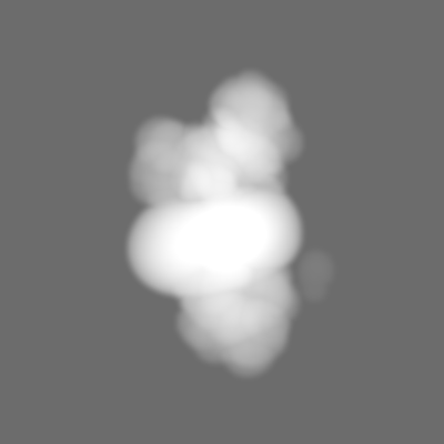
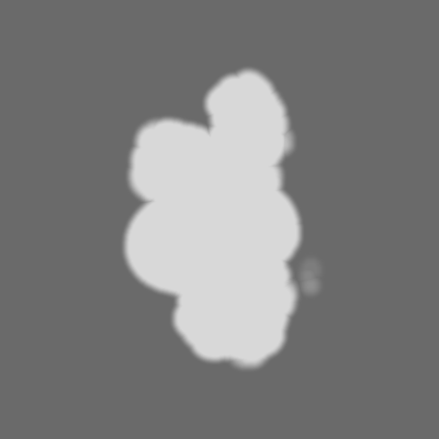
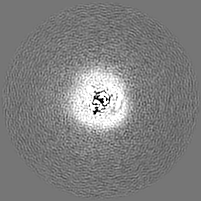
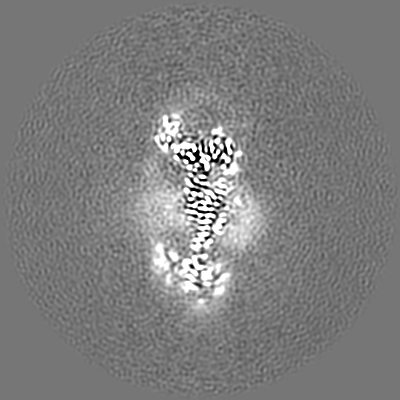
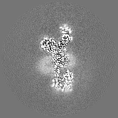
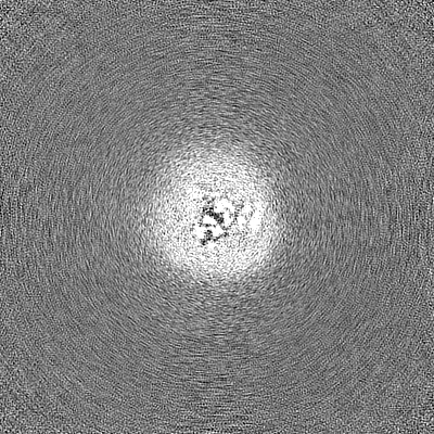
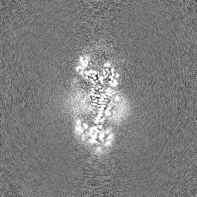
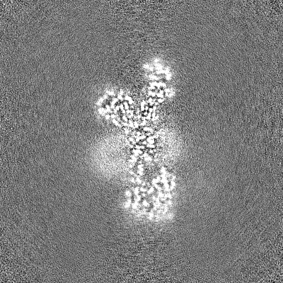
Human ER membrane protein complex (EMC) in GDN, 9-subunit map
マップデータ
Full map file
試料
複合体: Human ER Membrane Protein Complex
タンパク質・ペプチド: x 9種
リガンド: x 2種
キーワード
Insertase / endoplasmic reticulum / transmembrane chaperone / MEMBRANE PROTEIN
機能・相同性
機能・相同性情報
extrinsic component of endoplasmic reticulum membrane / inorganic cation transmembrane transporter activity / EMC complex / omegasome membrane / protein insertion into ER membrane by stop-transfer membrane-anchor sequence / magnesium ion transport / tail-anchored membrane protein insertion into ER membrane / Miscellaneous transport and binding events / cobalt ion transmembrane transporter activity / ferrous iron transmembrane transporter activity ...extrinsic component of endoplasmic reticulum membrane / inorganic cation transmembrane transporter activity / EMC complex / omegasome membrane / protein insertion into ER membrane by stop-transfer membrane-anchor sequence / magnesium ion transport / tail-anchored membrane protein insertion into ER membrane / Miscellaneous transport and binding events / cobalt ion transmembrane transporter activity / ferrous iron transmembrane transporter activity / copper ion transport / magnesium ion transmembrane transporter activity / autophagosome assembly / RHOA GTPase cycle / positive regulation of endothelial cell proliferation / positive regulation of endothelial cell migration / positive regulation of angiogenesis / carbohydrate binding / early endosome membrane / angiogenesis / early endosome / Golgi membrane / apoptotic process / endoplasmic reticulum membrane / endoplasmic reticulum / Golgi apparatus / protein-containing complex / extracellular region / membrane / plasma membrane / cytoplasm 類似検索 - 分子機能
EMC1 N-terminal beta-propeller domain / ER membrane protein complex subunit 8/9 / : / Uncharacterised protein family (UPF0172) / EMC2 TPR-like repeat domain / TMEM85/ER membrane protein complex subunit 4 / ER membrane protein complex subunit 4 / ER membrane protein complex subunit 7, beta-sandwich domain / ER membrane protein complex subunit 7 / ER membrane protein complex subunit 7, beta-sandwich domain ...EMC1 N-terminal beta-propeller domain / ER membrane protein complex subunit 8/9 / : / Uncharacterised protein family (UPF0172) / EMC2 TPR-like repeat domain / TMEM85/ER membrane protein complex subunit 4 / ER membrane protein complex subunit 4 / ER membrane protein complex subunit 7, beta-sandwich domain / ER membrane protein complex subunit 7 / ER membrane protein complex subunit 7, beta-sandwich domain / ER membrane protein complex subunit 6 / ER membrane protein complex subunit 3 / ER membrane protein complex subunit 1, C-terminal / Membrane magnesium transporter / ER membrane protein complex subunit 1 / ER membrane protein complex subunit 6-like / EMC6 / ER membrane protein complex subunit 1, C-terminal / Membrane magnesium transporter / ER membrane protein complex subunit 10 / ER membrane protein complex subunit 2-like / Integral membrane protein EMC3/TMCO1-like / Integral membrane protein EMC3/TMCO1-like / Integral membrane protein DUF106 / Carbohydrate-binding-like fold / Quinoprotein alcohol dehydrogenase-like superfamily / TPR repeat region circular profile. / TPR repeat profile. / MPN domain / MPN domain profile. / Tetratricopeptide repeats / Tetratricopeptide repeat / Tetratricopeptide-like helical domain superfamily / WD40/YVTN repeat-like-containing domain superfamily 類似検索 - ドメイン・相同性
ER membrane protein complex subunit 8 / ER membrane protein complex subunit 2 / ER membrane protein complex subunit 4 / ER membrane protein complex subunit 10 / ER membrane protein complex subunit 5 / ER membrane protein complex subunit 1 / ER membrane protein complex subunit 6 / Endoplasmic reticulum membrane protein complex subunit 7 / ER membrane protein complex subunit 3 類似検索 - 構成要素
National Institutes of Health/National Institute of General Medical Sciences (NIH/NIGMS)
DP2GM137412
米国
引用
ジャーナル: J Cell Biol / 年: 2023 タイトル: A selectivity filter in the ER membrane protein complex limits protein misinsertion at the ER. 著者: Tino Pleiner / Masami Hazu / Giovani Pinton Tomaleri / Vy N Nguyen / Kurt Januszyk / Rebecca M Voorhees / 要旨: Tail-anchored (TA) proteins play essential roles in mammalian cells, and their accurate localization is critical for proteostasis. Biophysical similarities lead to mistargeting of mitochondrial TA ...Tail-anchored (TA) proteins play essential roles in mammalian cells, and their accurate localization is critical for proteostasis. Biophysical similarities lead to mistargeting of mitochondrial TA proteins to the ER, where they are delivered to the insertase, the ER membrane protein complex (EMC). Leveraging an improved structural model of the human EMC, we used mutagenesis and site-specific crosslinking to map the path of a TA protein from its cytosolic capture by methionine-rich loops to its membrane insertion through a hydrophilic vestibule. Positively charged residues at the entrance to the vestibule function as a selectivity filter that uses charge-repulsion to reject mitochondrial TA proteins. Similarly, this selectivity filter retains the positively charged soluble domains of multipass substrates in the cytosol, thereby ensuring they adopt the correct topology and enforcing the "positive-inside" rule. Substrate discrimination by the EMC provides a biochemical explanation for one role of charge in TA protein sorting and protects compartment integrity by limiting protein misinsertion.
 ムービー
ムービー コントローラー


 データを開く
 基本情報
基本情報
 マップデータ
 試料
試料 キーワード
キーワード 機能・相同性情報
 Homo sapiens (ヒト)
 データ登録者
 米国, 1件
  引用
引用 構造の表示
構造の表示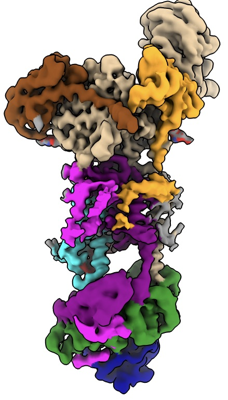
 ダウンロードとリンク
 emd_40245.png
 http://ftp.pdbj.org/pub/emdb/structures/EMD-40245


 Z (Sec.)
 Y (Row.)
 X (Col.)


 試料の構成要素

 解析
解析 電子顕微鏡法
 FIELD EMISSION GUN