 Movie
Movie Controller


+ Open data

- Basic information
| Entry | Database: PDB / ID: 8s9s | ||||||
|---|---|---|---|---|---|---|---|
| Title | Structure of the human ER membrane protein complex (EMC) in GDN | ||||||
 Components Components |
| ||||||
 Keywords Keywords | MEMBRANE PROTEIN / Insertase / endoplasmic reticulum / transmembrane chaperone | ||||||
| Function / homology |  Function and homology information Function and homology informationextrinsic component of endoplasmic reticulum membrane / EMC complex / : / omegasome membrane / protein insertion into ER membrane by stop-transfer membrane-anchor sequence / magnesium ion transport / tail-anchored membrane protein insertion into ER membrane / Miscellaneous transport and binding events / cobalt ion transmembrane transporter activity / ferrous iron transmembrane transporter activity ...extrinsic component of endoplasmic reticulum membrane / EMC complex / : / omegasome membrane / protein insertion into ER membrane by stop-transfer membrane-anchor sequence / magnesium ion transport / tail-anchored membrane protein insertion into ER membrane / Miscellaneous transport and binding events / cobalt ion transmembrane transporter activity / ferrous iron transmembrane transporter activity / copper ion transport / magnesium ion transmembrane transporter activity / RHOA GTPase cycle / autophagosome assembly / positive regulation of endothelial cell proliferation / positive regulation of endothelial cell migration / positive regulation of angiogenesis / carbohydrate binding / angiogenesis / early endosome membrane / early endosome / Golgi membrane / apoptotic process / endoplasmic reticulum membrane / endoplasmic reticulum / Golgi apparatus / protein-containing complex / extracellular region / membrane / plasma membrane / cytoplasm Similarity search - Function | ||||||
| Biological species |  Homo sapiens (human) Homo sapiens (human) | ||||||
| Method | ELECTRON MICROSCOPY / single particle reconstruction / cryo EM / Resolution: 3.6 Å | ||||||
 Authors Authors | Tomaleri, G.P. / Nguyen, V.N. / Voorhees, R.M. | ||||||
| Funding support |  United States, 1items United States, 1items
| ||||||
 Citation Citation | Journal: J Cell Biol / Year: 2023 Title: A selectivity filter in the ER membrane protein complex limits protein misinsertion at the ER. Authors: Tino Pleiner / Masami Hazu / Giovani Pinton Tomaleri / Vy N Nguyen / Kurt Januszyk / Rebecca M Voorhees / Abstract: Tail-anchored (TA) proteins play essential roles in mammalian cells, and their accurate localization is critical for proteostasis. Biophysical similarities lead to mistargeting of mitochondrial TA ...Tail-anchored (TA) proteins play essential roles in mammalian cells, and their accurate localization is critical for proteostasis. Biophysical similarities lead to mistargeting of mitochondrial TA proteins to the ER, where they are delivered to the insertase, the ER membrane protein complex (EMC). Leveraging an improved structural model of the human EMC, we used mutagenesis and site-specific crosslinking to map the path of a TA protein from its cytosolic capture by methionine-rich loops to its membrane insertion through a hydrophilic vestibule. Positively charged residues at the entrance to the vestibule function as a selectivity filter that uses charge-repulsion to reject mitochondrial TA proteins. Similarly, this selectivity filter retains the positively charged soluble domains of multipass substrates in the cytosol, thereby ensuring they adopt the correct topology and enforcing the "positive-inside" rule. Substrate discrimination by the EMC provides a biochemical explanation for one role of charge in TA protein sorting and protects compartment integrity by limiting protein misinsertion. | ||||||
| History |
|
- Structure visualization
| Structure viewer | Molecule: MolmilJmol/JSmol |
|---|
- Downloads & links
-Download
| PDBx/mmCIF format | 8s9s.cif.gz | 500.3 KB | Display | PDBx/mmCIF format |
|---|---|---|---|---|
| PDB format | pdb8s9s.ent.gz | Display | PDB format | |
| PDBx/mmJSON format | 8s9s.json.gz | Tree view | PDBx/mmJSON format | |
| Others |  Other downloads Other downloads |
-Validation report
| Arichive directory | https://data.pdbj.org/pub/pdb/validation_reports/s9/8s9sftp://data.pdbj.org/pub/pdb/validation_reports/s9/8s9s | HTTPS FTP |
|---|
-Related structure data
| Related structure data |  40245MC M: map data used to model this data C: citing same article ( |
|---|---|
| Similar structure data |


-Links
 PDBj
PDBj


- Assembly
| Deposited unit | 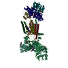
|
|---|---|
| 1 |
|
-Components
-ER membrane protein complex subunit ... , 8 types, 8 molecules 123467810
| #1: Protein | Mass: 111886.141 Da / Num. of mol.: 1 / Source method: isolated from a natural source / Source: (natural) Homo sapiens (human) / References: UniProt: Q8N766 |
|---|---|
| #2: Protein | Mass: 34882.531 Da / Num. of mol.: 1 / Source method: isolated from a natural source / Source: (natural) Homo sapiens (human) / References: UniProt: Q15006 |
| #3: Protein | Mass: 29981.924 Da / Num. of mol.: 1 / Source method: isolated from a natural source / Source: (natural) Homo sapiens (human) / References: UniProt: Q9P0I2 |
| #4: Protein | Mass: 20104.572 Da / Num. of mol.: 1 / Source method: isolated from a natural source / Source: (natural) Homo sapiens (human) / References: UniProt: Q5J8M3 |
| #6: Protein | Mass: 12029.248 Da / Num. of mol.: 1 / Source method: isolated from a natural source / Source: (natural) Homo sapiens (human) / References: UniProt: Q9BV81 |
| #7: Protein | Mass: 26501.586 Da / Num. of mol.: 1 / Source method: isolated from a natural source / Source: (natural) Homo sapiens (human) / References: UniProt: Q9NPA0 |
| #8: Protein | Mass: 23807.076 Da / Num. of mol.: 1 / Source method: isolated from a natural source / Source: (natural) Homo sapiens (human) / References: UniProt: O43402 |
| #9: Protein | Mass: 27375.797 Da / Num. of mol.: 1 / Source method: isolated from a natural source / Source: (natural) Homo sapiens (human) / References: UniProt: Q5UCC4 |
-Protein / Non-polymers , 2 types, 7 molecules 5
| #12: Chemical | ChemComp-PCW /  Mass: 787.121 Da / Num. of mol.: 6 / Source method: obtained synthetically / Formula: C44H85NO8P / Comment: DOPC, phospholipid*YM Mass: 787.121 Da / Num. of mol.: 6 / Source method: obtained synthetically / Formula: C44H85NO8P / Comment: DOPC, phospholipid*YM#5: Protein | | Mass: 14706.786 Da / Num. of mol.: 1 / Source method: isolated from a natural source / Source: (natural) Homo sapiens (human) / References: UniProt: Q8N4V1 |
|---|
-Sugars , 2 types, 4 molecules 
| #10: Polysaccharide | Source method: isolated from a genetically manipulated source #11: Sugar | ChemComp-NAG / |  Type: D-saccharide, beta linking / Mass: 221.208 Da / Num. of mol.: 1 / Source method: obtained synthetically / Formula: C8H15NO6 Type: D-saccharide, beta linking / Mass: 221.208 Da / Num. of mol.: 1 / Source method: obtained synthetically / Formula: C8H15NO6 |
|---|
-Details
| Has ligand of interest | N |
|---|---|
| Has protein modification | Y |
-Experimental details
-Experiment
| Experiment | Method: ELECTRON MICROSCOPY |
|---|---|
| EM experiment | Aggregation state: PARTICLE / 3D reconstruction method: single particle reconstruction |
- Sample preparation
| Component | Name: Human ER Membrane Protein Complex / Type: COMPLEX / Entity ID: #1-#9 / Source: NATURAL | ||||||||||||||||||||||||||||||
|---|---|---|---|---|---|---|---|---|---|---|---|---|---|---|---|---|---|---|---|---|---|---|---|---|---|---|---|---|---|---|---|
| Molecular weight | Experimental value: NO | ||||||||||||||||||||||||||||||
| Source (natural) | Organism: Homo sapiens (human) / Cell: HEK293 | ||||||||||||||||||||||||||||||
| Buffer solution | pH: 7.5 | ||||||||||||||||||||||||||||||
| Buffer component |
| ||||||||||||||||||||||||||||||
| Specimen | Conc.: 0.5 mg/ml / Embedding applied: NO / Shadowing applied: NO / Staining applied: NO / Vitrification applied: YES / Details: Sample solubilized and purified in GDN | ||||||||||||||||||||||||||||||
| Specimen support | Grid material: GOLD / Grid mesh size: 300 divisions/in. / Grid type: Quantifoil R1.2/1.3 | ||||||||||||||||||||||||||||||
| Vitrification | Instrument: FEI VITROBOT MARK IV / Cryogen name: ETHANE / Humidity: 95 % / Chamber temperature: 279 K |
- Electron microscopy imaging
| Experimental equipment |  Model: Titan Krios / Image courtesy: FEI Company |
|---|---|
| Microscopy | Model: FEI TITAN KRIOS |
| Electron gun | Electron source:  FIELD EMISSION GUN / Accelerating voltage: 300 kV / Illumination mode: SPOT SCAN FIELD EMISSION GUN / Accelerating voltage: 300 kV / Illumination mode: SPOT SCAN |
| Electron lens | Mode: DARK FIELD / Nominal magnification: 105000 X / Nominal defocus max: 3000 nm / Nominal defocus min: 1000 nm / Cs: 2.7 mm / Alignment procedure: COMA FREE |
| Specimen holder | Cryogen: NITROGEN / Specimen holder model: FEI TITAN KRIOS AUTOGRID HOLDER |
| Image recording | Average exposure time: 2.66 sec. / Electron dose: 60 e/Å2 / Film or detector model: GATAN K3 (6k x 4k) / Num. of grids imaged: 2 / Num. of real images: 11822 |
| EM imaging optics | Energyfilter name: GIF Quantum LS / Energyfilter slit width: 20 eV |
- Processing
| Software |
| |||||||||||||||||||||||||||||||||||||||||||||
|---|---|---|---|---|---|---|---|---|---|---|---|---|---|---|---|---|---|---|---|---|---|---|---|---|---|---|---|---|---|---|---|---|---|---|---|---|---|---|---|---|---|---|---|---|---|---|
| EM software |
| |||||||||||||||||||||||||||||||||||||||||||||
| CTF correction | Type: PHASE FLIPPING AND AMPLITUDE CORRECTION | |||||||||||||||||||||||||||||||||||||||||||||
| Particle selection | Num. of particles selected: 1034250 | |||||||||||||||||||||||||||||||||||||||||||||
| 3D reconstruction | Resolution: 3.6 Å / Resolution method: FSC 0.143 CUT-OFF / Num. of particles: 156706 / Symmetry type: POINT | |||||||||||||||||||||||||||||||||||||||||||||
| Atomic model building | Protocol: RIGID BODY FIT / Space: REAL | |||||||||||||||||||||||||||||||||||||||||||||
| Refinement | Cross valid method: NONE Stereochemistry target values: GeoStd + Monomer Library + CDL v1.2 | |||||||||||||||||||||||||||||||||||||||||||||
| Displacement parameters | Biso mean: 115.7 Å2 | |||||||||||||||||||||||||||||||||||||||||||||
| Refine LS restraints |
|
