ムービー
ムービー コントローラー


+ データを開く

- 基本情報
基本情報
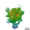
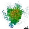
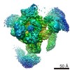
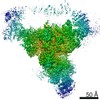
| 登録情報 | データベース: EMDB / ID: EMD-3979 | |||||||||
|---|---|---|---|---|---|---|---|---|---|---|
| タイトル | Post-catalytic P complex spliceosome with 3' splice site docked | |||||||||
 マップデータ マップデータ | ||||||||||
 試料 試料 |
| |||||||||
| 機能・相同性 |  機能・相同性情報 機能・相同性情報U2-type post-spliceosomal complex / U2-type post-mRNA release spliceosomal complex / mRNA branch site recognition / spliceosomal complex disassembly / cellular bud site selection / pre-mRNA 3'-splice site binding / post-mRNA release spliceosomal complex / generation of catalytic spliceosome for first transesterification step / nuclear mRNA surveillance / cis assembly of pre-catalytic spliceosome ...U2-type post-spliceosomal complex / U2-type post-mRNA release spliceosomal complex / mRNA branch site recognition / spliceosomal complex disassembly / cellular bud site selection / pre-mRNA 3'-splice site binding / post-mRNA release spliceosomal complex / generation of catalytic spliceosome for first transesterification step / nuclear mRNA surveillance / cis assembly of pre-catalytic spliceosome / splicing factor binding / spliceosome conformational change to release U4 (or U4atac) and U1 (or U11) / U4/U6 snRNP / Prp19 complex / 7-methylguanosine cap hypermethylation / pICln-Sm protein complex / pre-mRNA binding / U2-type catalytic step 1 spliceosome / small nuclear ribonucleoprotein complex / SMN-Sm protein complex / spliceosomal tri-snRNP complex / U2-type spliceosomal complex / mRNA cis splicing, via spliceosome / commitment complex / U2-type prespliceosome assembly / poly(U) RNA binding / U2-type catalytic step 2 spliceosome / U4 snRNP / U2 snRNP / U1 snRNP / U2-type prespliceosome / precatalytic spliceosome / Formation of TC-NER Pre-Incision Complex / generation of catalytic spliceosome for second transesterification step / spliceosomal complex assembly / Gap-filling DNA repair synthesis and ligation in TC-NER / DNA replication origin binding / mRNA 5'-splice site recognition / protein K63-linked ubiquitination / regulation of RNA splicing / mRNA 3'-splice site recognition / Dual incision in TC-NER / spliceosomal tri-snRNP complex assembly / DNA replication initiation / U5 snRNA binding / U5 snRNP / U2 snRNA binding / U6 snRNA binding / spliceosomal snRNP assembly / positive regulation of cell cycle / pre-mRNA intronic binding / U1 snRNA binding / U4/U6 x U5 tri-snRNP complex / catalytic step 2 spliceosome / RNA splicing / nuclear periphery / positive regulation of RNA splicing / RNA polymerase II transcription regulatory region sequence-specific DNA binding / spliceosomal complex / RING-type E3 ubiquitin transferase / mRNA splicing, via spliceosome / ubiquitin-protein transferase activity / metallopeptidase activity / ubiquitin protein ligase activity / RNA helicase activity / DNA-binding transcription factor activity, RNA polymerase II-specific / RNA helicase / cell cycle / DNA repair / GTPase activity / mRNA binding / chromatin binding / chromatin / GTP binding / ATP hydrolysis activity / mitochondrion / RNA binding / zinc ion binding / ATP binding / identical protein binding / nucleus / metal ion binding / cytoplasm / cytosol 類似検索 - 分子機能 | |||||||||
| 生物種 |  | |||||||||
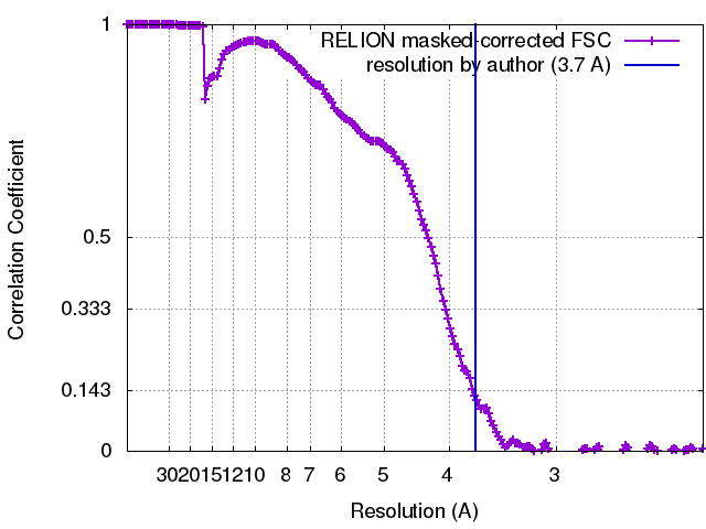
| 手法 | 単粒子再構成法 / クライオ電子顕微鏡法 / 解像度: 3.7 Å | |||||||||
 データ登録者 データ登録者 | Wilkinson ME / Fica SM / Galej WP / Norman CM / Newman AJ / Nagai K | |||||||||
| 資金援助 |  英国, 2件 英国, 2件
| |||||||||
 引用 引用 | ジャーナル: Science / 年: 2017 タイトル: Postcatalytic spliceosome structure reveals mechanism of 3'-splice site selection. 著者: Max E Wilkinson / Sebastian M Fica / Wojciech P Galej / Christine M Norman / Andrew J Newman / Kiyoshi Nagai / 要旨: Introns are removed from eukaryotic messenger RNA precursors by the spliceosome in two transesterification reactions-branching and exon ligation. The mechanism of 3'-splice site recognition during ...Introns are removed from eukaryotic messenger RNA precursors by the spliceosome in two transesterification reactions-branching and exon ligation. The mechanism of 3'-splice site recognition during exon ligation has remained unclear. Here we present the 3.7-angstrom cryo-electron microscopy structure of the yeast P-complex spliceosome immediately after exon ligation. The 3'-splice site AG dinucleotide is recognized through non-Watson-Crick pairing with the 5' splice site and the branch-point adenosine. After the branching reaction, protein factors work together to remodel the spliceosome and stabilize a conformation competent for 3'-splice site docking, thereby promoting exon ligation. The structure accounts for the strict conservation of the GU and AG dinucleotides at the 5' and 3' ends of introns and provides insight into the catalytic mechanism of exon ligation. | |||||||||
| 履歴 |
|
- 構造の表示
構造の表示
| ムービー |
ムービービューア |
|---|---|
| 構造ビューア | EMマップ: SurfViewMolmilJmol/JSmol |
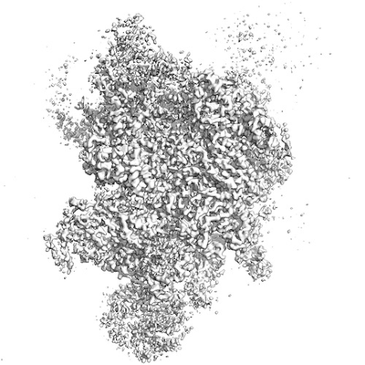
| 添付画像 |
- ダウンロードとリンク
-EMDBアーカイブ
| マップデータ | emd_3979.map.gz | 390.9 MB | EMDBマップデータ形式 | |
|---|---|---|---|---|
| ヘッダ (付随情報) | emd-3979-v30.xmlemd-3979.xml | 63.2 KB 63.2 KB | 表示 表示 | EMDBヘッダ |
| FSC (解像度算出) | emd_3979_fsc.xml | 17 KB | 表示 | FSCデータファイル |
| 画像 |  emd_3979.png emd_3979.png | 71 KB | ||
| アーカイブディレクトリ |  http://ftp.pdbj.org/pub/emdb/structures/EMD-3979ftp://ftp.pdbj.org/pub/emdb/structures/EMD-3979 http://ftp.pdbj.org/pub/emdb/structures/EMD-3979ftp://ftp.pdbj.org/pub/emdb/structures/EMD-3979 | HTTPS FTP |
-検証レポート
| 文書・要旨 | emd_3979_validation.pdf.gz | 323.3 KB | 表示 | EMDB検証レポート |
|---|---|---|---|---|
| 文書・詳細版 | emd_3979_full_validation.pdf.gz | 322.4 KB | 表示 | |
| XML形式データ | emd_3979_validation.xml.gz | 15.1 KB | 表示 | |
| アーカイブディレクトリ | https://ftp.pdbj.org/pub/emdb/validation_reports/EMD-3979ftp://ftp.pdbj.org/pub/emdb/validation_reports/EMD-3979 | HTTPS FTP |
-関連構造データ

-リンク
| EMDBのページ | EMDB (EBI/PDBe) / EMDataResource |
|---|---|
| 「今月の分子」の関連する項目 |


-マップ
| ファイル | ダウンロード / ファイル: emd_3979.map.gz / 形式: CCP4 / 大きさ: 421.9 MB / タイプ: IMAGE STORED AS FLOATING POINT NUMBER (4 BYTES) | ||||||||||||||||||||||||||||||||||||||||||||||||||||||||||||
|---|---|---|---|---|---|---|---|---|---|---|---|---|---|---|---|---|---|---|---|---|---|---|---|---|---|---|---|---|---|---|---|---|---|---|---|---|---|---|---|---|---|---|---|---|---|---|---|---|---|---|---|---|---|---|---|---|---|---|---|---|---|
| ボクセルのサイズ | X=Y=Z: 1.12 Å | ||||||||||||||||||||||||||||||||||||||||||||||||||||||||||||
| 密度 |
| ||||||||||||||||||||||||||||||||||||||||||||||||||||||||||||
| 対称性 | 空間群: 1 | ||||||||||||||||||||||||||||||||||||||||||||||||||||||||||||
| 詳細 | EMDB XML:
CCP4マップ ヘッダ情報:
| ||||||||||||||||||||||||||||||||||||||||||||||||||||||||||||
-添付データ
- 試料の構成要素
+全体 : P complex spliceosome with 3' exon docked
+超分子 #1: P complex spliceosome with 3' exon docked
+超分子 #2: P complex spliceosome
+超分子 #3: 3' exon
+分子 #1: U2 snRNA
+分子 #2: U5 snRNA
+分子 #3: U6 snRNA
+分子 #7: Ligated exons: UBC4 mRNA
+分子 #9: Intron lariat: UBC4 RNA
+分子 #4: Pre-mRNA-splicing factor Prp8
+分子 #5: Pre-mRNA-splicing factor SNU114
+分子 #6: Pre-mRNA-splicing factor CWC16
+分子 #8: Pre-mRNA-splicing factor CWC22
+分子 #10: Pre-mRNA-splicing factor PRP46
+分子 #11: Pre-mRNA-processing protein 45
+分子 #12: Pre-mRNA-splicing factor BUD31
+分子 #13: Pre-mRNA-splicing factor CWC2
+分子 #14: Pre-mRNA-splicing factor SLT11
+分子 #15: Pre-mRNA-splicing factor CEF1
+分子 #16: Pre-mRNA-splicing factor CWC15
+分子 #17: Pre-mRNA-splicing factor CWC21
+分子 #18: Pre-mRNA-splicing factor CLF1
+分子 #19: Pre-mRNA-splicing factor SYF1
+分子 #20: Pre-mRNA-splicing factor ATP-dependent RNA helicase PRP22
+分子 #21: U2 small nuclear ribonucleoprotein A'
+分子 #22: Unassigned structure
+分子 #23: U2 small nuclear ribonucleoprotein B''
+分子 #24: Pre-mRNA-splicing factor Prp18
+分子 #25: Small nuclear ribonucleoprotein-associated protein B
+分子 #26: Pre-mRNA-splicing factor SLU7
+分子 #27: Small nuclear ribonucleoprotein Sm D3
+分子 #28: Small nuclear ribonucleoprotein E
+分子 #29: Small nuclear ribonucleoprotein F
+分子 #30: Small nuclear ribonucleoprotein G
+分子 #31: Small nuclear ribonucleoprotein Sm D1
+分子 #32: Small nuclear ribonucleoprotein Sm D2
+分子 #33: Pre-mRNA-processing factor Prp17
+分子 #34: Pre-mRNA-splicing factor SNT309
+分子 #35: Pre-mRNA-processing factor Prp19
+分子 #36: Pre-mRNA-splicing factor SYF2
+分子 #37: MAGNESIUM ION
+分子 #38: INOSITOL HEXAKISPHOSPHATE

+分子 #39: GUANOSINE-5'-TRIPHOSPHATE

+分子 #40: ZINC ION
-実験情報
-構造解析
| 手法 | クライオ電子顕微鏡法 |
|---|---|
 解析 解析 | 単粒子再構成法 |
| 試料の集合状態 | particle |
-試料調製
| 濃度 | 1.4 mg/mL | ||||||||||||
|---|---|---|---|---|---|---|---|---|---|---|---|---|---|
| 緩衝液 | pH: 7.9 構成要素:
| ||||||||||||
| グリッド | モデル: Quantifoil R2/2 / 材質: COPPER / メッシュ: 400 / 支持フィルム - 材質: CARBON / 支持フィルム - トポロジー: CONTINUOUS / 支持フィルム - Film thickness: 7.0 nm / 前処理 - タイプ: GLOW DISCHARGE / 前処理 - 雰囲気: AIR | ||||||||||||
| 凍結 | 凍結剤: ETHANE / チャンバー内湿度: 100 % / チャンバー内温度: 277 K / 装置: FEI VITROBOT MARK III 詳細: 3 uL sample was applied to the grid, left for 30s, then blotted for 3s and immediately plunged into liquid ethane.. |
- 電子顕微鏡法
| 顕微鏡 | FEI TITAN KRIOS |
|---|---|
| 撮影 | フィルム・検出器のモデル: GATAN K2 SUMMIT (4k x 4k) 検出モード: COUNTING / デジタル化 - 画像ごとのフレーム数: 1-20 / 撮影したグリッド数: 1 / 実像数: 2295 / 平均露光時間: 12.0 sec. / 平均電子線量: 47.0 e/Å2 |
| 電子線 | 加速電圧: 300 kV / 電子線源:  FIELD EMISSION GUN FIELD EMISSION GUN |
| 電子光学系 | C2レンズ絞り径: 50.0 µm / 最大 デフォーカス(補正後): 3.0 µm / 最小 デフォーカス(補正後): 0.2 µm / 照射モード: FLOOD BEAM / 撮影モード: BRIGHT FIELD / Cs: 2.7 mm / 倍率(公称値): 105000 |
| 試料ステージ | 試料ホルダーモデル: FEI TITAN KRIOS AUTOGRID HOLDER ホルダー冷却材: NITROGEN |
| 実験機器 |  モデル: Titan Krios / 画像提供: FEI Company |