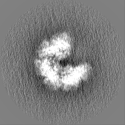
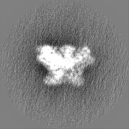
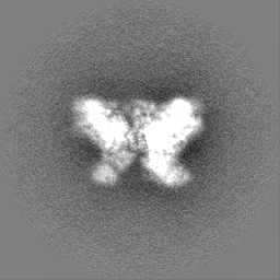
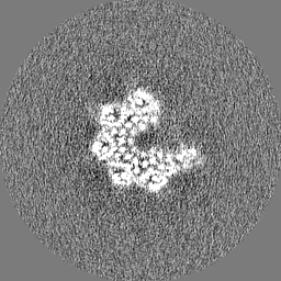
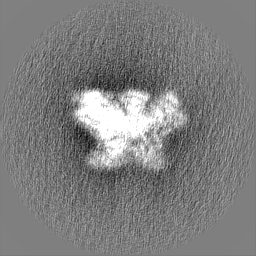
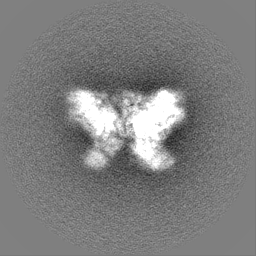
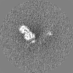
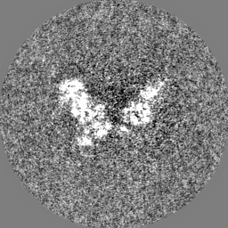
- EMDB-36450: cryo-EM structure of a CED-4 hexamer -
+
Open data
ID or keywords:
Loading...
-
Basic information
Entry
Database: EMDB / ID: EMD-36450
Title
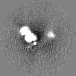
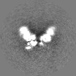
cryo-EM structure of a CED-4 hexamer
Map data
Sample
Complex: CED-4 hexamer
Protein or peptide: Cell death protein 4
Ligand: MAGNESIUM ION
Ligand: ADENOSINE-5'-TRIPHOSPHATE
Keywords
CED-4 apoptosome / Hexamer / APOPTOSIS
Function / homology
Function and homology information
BH1 domain binding / regulation of development, heterochronic / positive regulation of apoptotic process involved in development / caspase complex / positive regulation of synapse pruning / peptidase activator activity involved in apoptotic process / caspase binding / positive regulation of protein processing / embryonic morphogenesis / apoptotic process involved in development ...BH1 domain binding / regulation of development, heterochronic / positive regulation of apoptotic process involved in development / caspase complex / positive regulation of synapse pruning / peptidase activator activity involved in apoptotic process / caspase binding / positive regulation of protein processing / embryonic morphogenesis / apoptotic process involved in development / cysteine-type endopeptidase activator activity / actin filament depolymerization / embryo development ending in birth or egg hatching / negative regulation of execution phase of apoptosis / muscle cell cellular homeostasis / regulation of cell size / cysteine-type endopeptidase activator activity involved in apoptotic process / BH3 domain binding / regulation of cell adhesion / endopeptidase activator activity / regulation of protein stability / ADP binding / defense response to Gram-negative bacterium / positive regulation of apoptotic process / apoptotic process / negative regulation of apoptotic process / perinuclear region of cytoplasm / magnesium ion binding / protein-containing complex / mitochondrion / ATP binding / membrane / identical protein binding / nucleus / cytosol Similarity search - Function
Journal: Life Sci Alliance / Year: 2023 Title: Structural insights into CED-3 activation. Authors: Yini Li / Lu Tian / Ying Zhang / Yigong Shi / Abstract: In , onset of programmed cell death is marked with the activation of CED-3, a process that requires assembly of the CED-4 apoptosome. Activated CED-3 forms a holoenzyme with the CED-4 apoptosome to ...In , onset of programmed cell death is marked with the activation of CED-3, a process that requires assembly of the CED-4 apoptosome. Activated CED-3 forms a holoenzyme with the CED-4 apoptosome to cleave a wide range of substrates, leading to irreversible cell death. Despite decades of investigations, the underlying mechanism of CED-4-facilitated CED-3 activation remains elusive. Here, we report cryo-EM structures of the CED-4 apoptosome and three distinct CED-4/CED-3 complexes that mimic different activation stages for CED-3. In addition to the previously reported octamer in crystal structures, CED-4, alone or in complex with CED-3, exists in multiple oligomeric states. Supported by biochemical analyses, we show that the conserved CARD-CARD interaction promotes CED-3 activation, and initiation of programmed cell death is regulated by the dynamic organization of the CED-4 apoptosome.
In the structure databanks used in Yorodumi, some data are registered as the other names, "COVID-19 virus" and "2019-nCoV". Here are the details of the virus and the list of structure data.
Jan 31, 2019. EMDB accession codes are about to change! (news from PDBe EMDB page)
EMDB accession codes are about to change! (news from PDBe EMDB page)
The allocation of 4 digits for EMDB accession codes will soon come to an end. Whilst these codes will remain in use, new EMDB accession codes will include an additional digit and will expand incrementally as the available range of codes is exhausted. The current 4-digit format prefixed with “EMD-” (i.e. EMD-XXXX) will advance to a 5-digit format (i.e. EMD-XXXXX), and so on. It is currently estimated that the 4-digit codes will be depleted around Spring 2019, at which point the 5-digit format will come into force.
The EM Navigator/Yorodumi systems omit the EMD- prefix.
Related info.:Q: What is EMD? / ID/Accession-code notation in Yorodumi/EM Navigator
Yorodumi is a browser for structure data from EMDB, PDB, SASBDB, etc.
This page is also the successor to EM Navigator detail page, and also detail information page/front-end page for Omokage search.
The word "yorodu" (or yorozu) is an old Japanese word meaning "ten thousand". "mi" (miru) is to see.
Related info.:EMDB / PDB / SASBDB / Comparison of 3 databanks / Yorodumi Search / Aug 31, 2016. New EM Navigator & Yorodumi / Yorodumi Papers / Jmol/JSmol / Function and homology information / Changes in new EM Navigator and Yorodumi
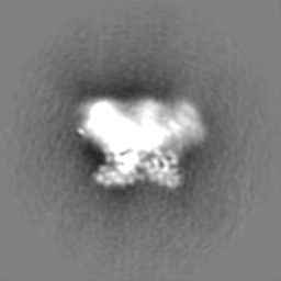
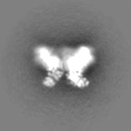
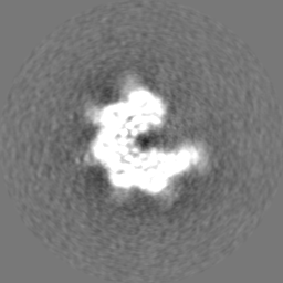
 Movie
Movie Controller


 Open data
 Basic information
 Map data
 Sample
 Keywords
 Function and homology information
 Authors
 China, 1 items
  Citation
 Structure visualization
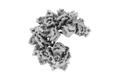
 Downloads & links
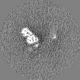
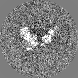
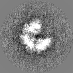
 emd_36450.png
 http://ftp.pdbj.org/pub/emdb/structures/EMD-36450


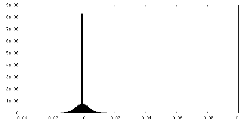
 Z (Sec.)
 Y (Row.)
 X (Col.)


 Sample components

 Processing
 Electron microscopy
 FIELD EMISSION GUN

