 Movie
Movie Controller


+ Open data

- Basic information
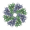
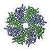
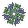
| Entry | Database: PDB / ID: 3lqq | ||||||
|---|---|---|---|---|---|---|---|
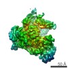
| Title | Structure of the CED-4 Apoptosome | ||||||
 Components Components | Cell death protein 4 | ||||||
 Keywords Keywords | APOPTOSIS / CED4 / apoptosome / Alternative splicing / ATP-binding / Mitochondrion / Nucleotide-binding | ||||||
| Function / homology |  Function and homology information Function and homology informationBH1 domain binding / regulation of development, heterochronic / positive regulation of apoptotic process involved in development / caspase complex / positive regulation of synapse pruning / peptidase activator activity involved in apoptotic process / positive regulation of protein processing / caspase binding / embryonic morphogenesis / apoptotic process involved in development ...BH1 domain binding / regulation of development, heterochronic / positive regulation of apoptotic process involved in development / caspase complex / positive regulation of synapse pruning / peptidase activator activity involved in apoptotic process / positive regulation of protein processing / caspase binding / embryonic morphogenesis / apoptotic process involved in development / cysteine-type endopeptidase activator activity / embryo development ending in birth or egg hatching / actin filament depolymerization / negative regulation of execution phase of apoptosis / muscle cell cellular homeostasis / regulation of cell size / cysteine-type endopeptidase activator activity involved in apoptotic process / BH3 domain binding / regulation of cell adhesion / endopeptidase activator activity / regulation of protein stability / ADP binding / defense response to Gram-negative bacterium / positive regulation of apoptotic process / apoptotic process / negative regulation of apoptotic process / perinuclear region of cytoplasm / magnesium ion binding / protein-containing complex / mitochondrion / ATP binding / membrane / identical protein binding / nucleus / cytosol Similarity search - Function | ||||||
| Biological species |  | ||||||
| Method |  X-RAY DIFFRACTION / SYNCHROTRON / MOLECULAR REPLACEMENT / Resolution: 3.534 Å X-RAY DIFFRACTION / SYNCHROTRON / MOLECULAR REPLACEMENT / Resolution: 3.534 Å | ||||||
 Authors Authors | Qi, S. / Pang, Y. / Shi, Y. / Yan, N. / Liu, Q. | ||||||
 Citation Citation | Journal: Cell(Cambridge,Mass.) / Year: 2010 Title: Crystal structure of the Caenorhabditis elegans apoptosome reveals an octameric assembly of CED-4. Authors: Qi, S. / Pang, Y. / Hu, Q. / Liu, Q. / Li, H. / Zhou, Y. / He, T. / Liang, Q. / Liu, Y. / Yuan, X. / Luo, G. / Li, H. / Wang, J. / Yan, N. / Shi, Y. | ||||||
| History |
|
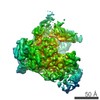
- Structure visualization
| Structure viewer | Molecule: MolmilJmol/JSmol |
|---|
- Downloads & links
-Download
| PDBx/mmCIF format | 3lqq.cif.gz | 215.7 KB | Display | PDBx/mmCIF format |
|---|---|---|---|---|
| PDB format | pdb3lqq.ent.gz | 171.3 KB | Display | PDB format |
| PDBx/mmJSON format | 3lqq.json.gz | Tree view | PDBx/mmJSON format | |
| Others |  Other downloads Other downloads |
-Validation report
| Arichive directory | https://data.pdbj.org/pub/pdb/validation_reports/lq/3lqqftp://data.pdbj.org/pub/pdb/validation_reports/lq/3lqq | HTTPS FTP |
|---|
-Related structure data
| Related structure data |  3lqrC  2a5yS S: Starting model for refinement C: citing same article ( |
|---|---|
| Similar structure data |

-Links
 PDBj
PDBj


- Assembly
| Deposited unit | 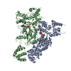
| |||||||||||||||||||||||||||||||||||||||||||||||||||||||||||||||||||||||||||||||||||||||||||||||||||||||||||||||||
|---|---|---|---|---|---|---|---|---|---|---|---|---|---|---|---|---|---|---|---|---|---|---|---|---|---|---|---|---|---|---|---|---|---|---|---|---|---|---|---|---|---|---|---|---|---|---|---|---|---|---|---|---|---|---|---|---|---|---|---|---|---|---|---|---|---|---|---|---|---|---|---|---|---|---|---|---|---|---|---|---|---|---|---|---|---|---|---|---|---|---|---|---|---|---|---|---|---|---|---|---|---|---|---|---|---|---|---|---|---|---|---|---|---|---|
| 1 | 
| |||||||||||||||||||||||||||||||||||||||||||||||||||||||||||||||||||||||||||||||||||||||||||||||||||||||||||||||||
| Unit cell |
| |||||||||||||||||||||||||||||||||||||||||||||||||||||||||||||||||||||||||||||||||||||||||||||||||||||||||||||||||
| Noncrystallographic symmetry (NCS) | NCS domain:
NCS domain segments:
|