 Movie
Movie Controller

[English] 日本語
 Yorodumi
- EMDB-31373: Cryo-EM structure of cyanobacterial phycobilisome from Synechococ... -
+ Open data

- Basic information
| Entry | Database: EMDB / ID: EMD-31373 | |||||||||
|---|---|---|---|---|---|---|---|---|---|---|
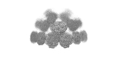
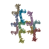
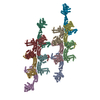
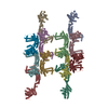
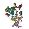
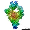
| Title | Cryo-EM structure of cyanobacterial phycobilisome from Synechococcus sp. PCC 7002 | |||||||||
 Map data Map data | ||||||||||
 Sample Sample |
| |||||||||
 Keywords Keywords | Phycobilisome / Synechococcus sp. PCC 7002 / Cryo-EM / photosynthesis | |||||||||
| Function / homology |  Function and homology information Function and homology informationLyases / phycobilisome / plasma membrane-derived thylakoid membrane / photosynthesis / lyase activity Similarity search - Function | |||||||||
| Biological species |  Synechococcus sp. PCC 7002 (bacteria) Synechococcus sp. PCC 7002 (bacteria) | |||||||||
| Method | single particle reconstruction / cryo EM / Resolution: 3.5 Å | |||||||||
 Authors Authors | Zheng L / Zheng Z | |||||||||
| Funding support | 1 items
| |||||||||
 Citation Citation | Journal: Nat Commun / Year: 2021 Title: Structural insight into the mechanism of energy transfer in cyanobacterial phycobilisomes. Authors: Lvqin Zheng / Zhenggao Zheng / Xiying Li / Guopeng Wang / Kun Zhang / Peijun Wei / Jindong Zhao / Ning Gao /  Abstract: Phycobilisomes (PBS) are the major light-harvesting machineries for photosynthesis in cyanobacteria and red algae and they have a hierarchical structure of a core and peripheral rods, with both ...Phycobilisomes (PBS) are the major light-harvesting machineries for photosynthesis in cyanobacteria and red algae and they have a hierarchical structure of a core and peripheral rods, with both consisting of phycobiliproteins and linker proteins. Here we report the cryo-EM structures of PBS from two cyanobacterial species, Anabaena 7120 and Synechococcus 7002. Both PBS are hemidiscoidal in shape and share a common triangular core structure. While the Anabaena PBS has two additional hexamers in the core linked by the 4th linker domain of ApcE (L). The PBS structures predict that, compared with the PBS from red algae, the cyanobacterial PBS could have more direct routes for energy transfer to ApcD. Structure-based systematic mutagenesis analysis of the chromophore environment of ApcD and ApcF subunits reveals that aromatic residues are critical to excitation energy transfer (EET). The structures also suggest that the linker protein could actively participate in the process of EET in both rods and the cores. These results provide insights into the organization of chromophores and the mechanisms of EET within cyanobacterial PBS. | |||||||||
| History |
|
- Structure visualization
| Movie |
Movie viewer |
|---|---|
| Structure viewer | EM map: SurfViewMolmilJmol/JSmol |
| Supplemental images |
- Downloads & links
-EMDB archive
| Map data | emd_31373.map.gz | 435.7 MB | EMDB map data format | |
|---|---|---|---|---|
| Header (meta data) | emd-31373-v30.xmlemd-31373.xml | 24.9 KB 24.9 KB | Display Display | EMDB header |
| Images |  emd_31373.png emd_31373.png | 36.4 KB | ||
| Filedesc metadata | emd-31373.cif.gz | 7.5 KB | ||
| Archive directory |  http://ftp.pdbj.org/pub/emdb/structures/EMD-31373ftp://ftp.pdbj.org/pub/emdb/structures/EMD-31373 http://ftp.pdbj.org/pub/emdb/structures/EMD-31373ftp://ftp.pdbj.org/pub/emdb/structures/EMD-31373 | HTTPS FTP |
-Related structure data
| Related structure data |  7extMC  7eydC M: atomic model generated by this map C: citing same article ( |
|---|---|
| Similar structure data |


-Links
| EMDB pages | EMDB (EBI/PDBe) / EMDataResource |
|---|
-Map
| File | Download / File: emd_31373.map.gz / Format: CCP4 / Size: 476.8 MB / Type: IMAGE STORED AS FLOATING POINT NUMBER (4 BYTES) | ||||||||||||||||||||||||||||||||||||||||||||||||||||||||||||||||||||
|---|---|---|---|---|---|---|---|---|---|---|---|---|---|---|---|---|---|---|---|---|---|---|---|---|---|---|---|---|---|---|---|---|---|---|---|---|---|---|---|---|---|---|---|---|---|---|---|---|---|---|---|---|---|---|---|---|---|---|---|---|---|---|---|---|---|---|---|---|---|
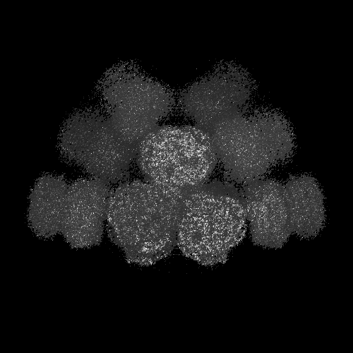
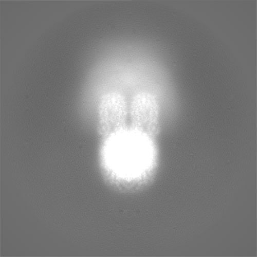
| Projections & slices | Image control
Images are generated by Spider. | ||||||||||||||||||||||||||||||||||||||||||||||||||||||||||||||||||||
| Voxel size | X=Y=Z: 1.055 Å | ||||||||||||||||||||||||||||||||||||||||||||||||||||||||||||||||||||
| Density |
| ||||||||||||||||||||||||||||||||||||||||||||||||||||||||||||||||||||
| Symmetry | Space group: 1 | ||||||||||||||||||||||||||||||||||||||||||||||||||||||||||||||||||||
| Details | EMDB XML:
CCP4 map header:
| ||||||||||||||||||||||||||||||||||||||||||||||||||||||||||||||||||||
 Z (Sec.)
 Y (Row.)
 X (Col.)


-Supplemental data
- Sample components
+Entire : Cryo-EM structure of cyanobacterial phycobilisome from Synechococ...
+Supramolecule #1: Cryo-EM structure of cyanobacterial phycobilisome from Synechococ...
+Macromolecule #1: Phycobilisome rod-core linker polypeptide CpcG
+Macromolecule #2: C-phycocyanin subunit alpha
+Macromolecule #3: C-phycocyanin subunit beta
+Macromolecule #4: Phycobilisome 32.3 kDa linker polypeptide, phycocyanin-associated, rod
+Macromolecule #5: Phycobiliprotein ApcE
+Macromolecule #6: Phycobilisome 7.8 kDa linker polypeptide, allophycocyanin-associa...
+Macromolecule #7: Allophycocyanin alpha subunit
+Macromolecule #8: Allophycocyanin beta subunit
+Macromolecule #9: Allophycocyanin subunit beta-18
+Macromolecule #10: Allophycocyanin subunit alpha-B
+Macromolecule #11: PHYCOCYANOBILIN

-Experimental details
-Structure determination
| Method | cryo EM |
|---|---|
 Processing Processing | single particle reconstruction |
| Aggregation state | particle |
-Sample preparation
| Buffer | pH: 7 |
|---|---|
| Vitrification | Cryogen name: ETHANE / Chamber humidity: 100 % / Instrument: FEI VITROBOT MARK IV |
- Electron microscopy
| Microscope | FEI TITAN KRIOS |
|---|---|
| Image recording | Film or detector model: GATAN K2 SUMMIT (4k x 4k) / Average electron dose: 64.0 e/Å2 |
| Electron beam | Acceleration voltage: 300 kV / Electron source:  FIELD EMISSION GUN FIELD EMISSION GUN |
| Electron optics | Illumination mode: FLOOD BEAM / Imaging mode: BRIGHT FIELD |
| Sample stage | Specimen holder model: FEI TITAN KRIOS AUTOGRID HOLDER / Cooling holder cryogen: NITROGEN |
| Experimental equipment |  Model: Titan Krios / Image courtesy: FEI Company |
