National Natural Science Foundation of China (NSFC)
32071199, 91940302
China
Chinese Academy of Sciences
XDB37010201
China
National Basic Research Program of China (973 Program)
2017YFA0504600
China
Citation
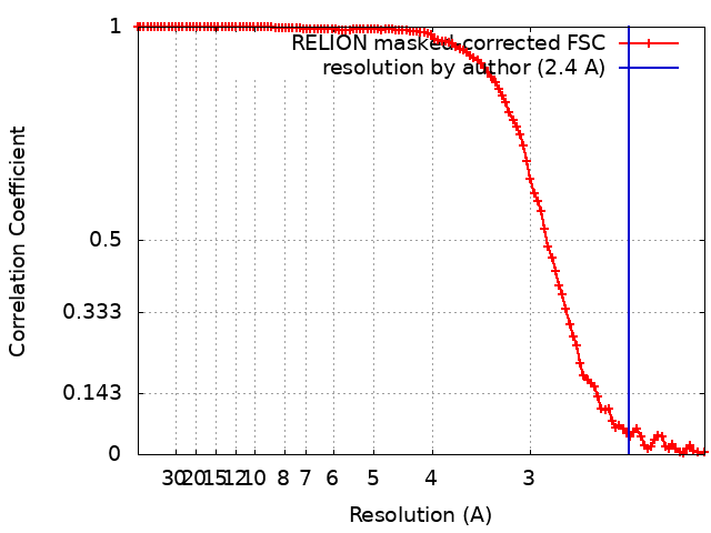
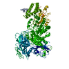
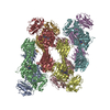
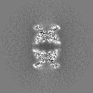
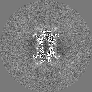
Journal: EMBO J / Year: 2021 Title: Molecular and structural mechanisms of ZZ domain-mediated cargo selection by Nbr1. Authors: Ying-Ying Wang / Jianxiu Zhang / Xiao-Man Liu / Yulu Li / Jianhua Sui / Meng-Qiu Dong / Keqiong Ye / Li-Lin Du / Abstract: In selective autophagy, cargo selectivity is determined by autophagy receptors. However, it remains scarcely understood how autophagy receptors recognize specific protein cargos. In the fission yeast ...In selective autophagy, cargo selectivity is determined by autophagy receptors. However, it remains scarcely understood how autophagy receptors recognize specific protein cargos. In the fission yeast Schizosaccharomyces pombe, a selective autophagy pathway termed Nbr1-mediated vacuolar targeting (NVT) employs Nbr1, an autophagy receptor conserved across eukaryotes including humans, to target cytosolic hydrolases into the vacuole. Here, we identify two new NVT cargos, the mannosidase Ams1 and the aminopeptidase Ape4, that bind competitively to the first ZZ domain of Nbr1 (Nbr1-ZZ1). High-resolution cryo-EM analyses reveal how a single ZZ domain recognizes two distinct protein cargos. Nbr1-ZZ1 not only recognizes the N-termini of cargos via a conserved acidic pocket, similar to other characterized ZZ domains, but also engages additional parts of cargos in a cargo-specific manner. Our findings unveil a single-domain bispecific mechanism of autophagy cargo recognition, elucidate its underlying structural basis, and expand the understanding of ZZ domain-mediated protein-protein interactions.
History
Deposition
Oct 28, 2020
-
Header (metadata) release
Jul 14, 2021
-
Map release
Jul 14, 2021
-
Update
May 29, 2024
-
Current status
May 29, 2024
Processing site: PDBj / Status: Released
-
Structure visualization
Movie
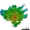
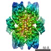
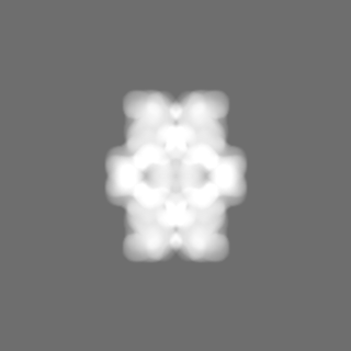
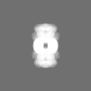
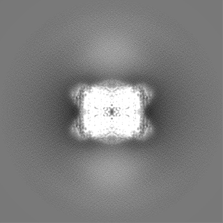
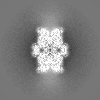
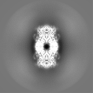
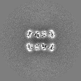
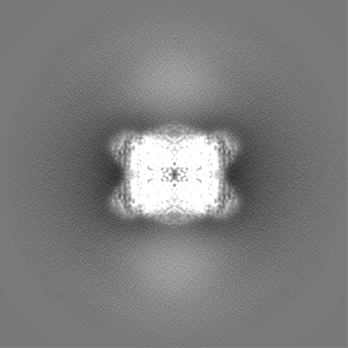
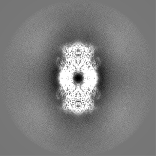
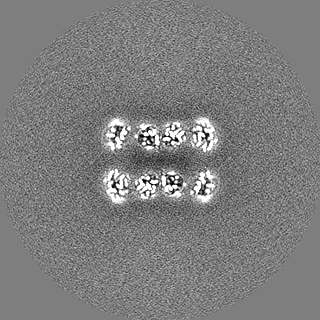
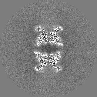
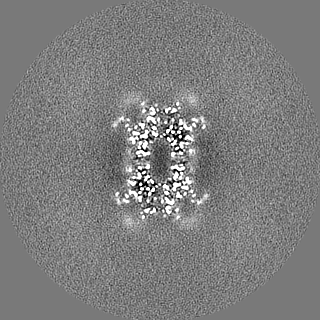
Surface view with section colored by density value
Protein or peptide: Alpha-mannosidase,ZZ-type zinc finger-containing protein P35G2.11c,Maltose/maltodextrin-binding periplasmic protein
Ligand: ZINC ION
Ligand: water
-
Supramolecule #1: Ams1 and Nbr1 fusion protein
Supramolecule
Name: Ams1 and Nbr1 fusion protein / type: complex / ID: 1 / Parent: 0 / Macromolecule list: #1 Details: The fusion protein comprises of the full-length Ams1, a linker sequence (GFKKASSSDNKEQ), residues 53-180 of Nbr1, and maltose binding protein (MBP).
Source (natural)
Organism: Schizosaccharomyces pombe 972h- (yeast)
Molecular weight
Theoretical: 520 KDa
-
Macromolecule #1: Alpha-mannosidase,ZZ-type zinc finger-containing protein P35G2.11...
Macromolecule
Name: Alpha-mannosidase,ZZ-type zinc finger-containing protein P35G2.11c,Maltose/maltodextrin-binding periplasmic protein type: protein_or_peptide / ID: 1 Details: The fusion protein comprises of the full-length Ams1, a linker sequence (GFKKASSSDNKEQ), residues 53-180 of Nbr1, and maltose binding protein (MBP). Number of copies: 4 / Enantiomer: LEVO / EC number: alpha-mannosidase
Model: Quantifoil R2/1 / Material: GOLD / Mesh: 300 / Support film - Material: CARBON / Support film - topology: HOLEY ARRAY / Pretreatment - Type: PLASMA CLEANING / Pretreatment - Time: 60 sec.
Vitrification
Cryogen name: ETHANE / Chamber humidity: 100 % / Chamber temperature: 277 K / Instrument: FEI VITROBOT MARK IV / Details: blot for 5 seconds before plunging.
-
Electron microscopy
Microscope
FEI TITAN KRIOS
Specialist optics
Energy filter - Name: GIF Tridiem 4K / Energy filter - Slit width: 20 eV
Image recording
Film or detector model: GATAN K2 SUMMIT (4k x 4k) / Detector mode: SUPER-RESOLUTION / Number real images: 2430 / Average exposure time: 8.9 sec. / Average electron dose: 60.0 e/Å2
Electron beam
Acceleration voltage: 300 kV / Electron source: FIELD EMISSION GUN
In the structure databanks used in Yorodumi, some data are registered as the other names, "COVID-19 virus" and "2019-nCoV". Here are the details of the virus and the list of structure data.
Jan 31, 2019. EMDB accession codes are about to change! (news from PDBe EMDB page)
EMDB accession codes are about to change! (news from PDBe EMDB page)
The allocation of 4 digits for EMDB accession codes will soon come to an end. Whilst these codes will remain in use, new EMDB accession codes will include an additional digit and will expand incrementally as the available range of codes is exhausted. The current 4-digit format prefixed with “EMD-” (i.e. EMD-XXXX) will advance to a 5-digit format (i.e. EMD-XXXXX), and so on. It is currently estimated that the 4-digit codes will be depleted around Spring 2019, at which point the 5-digit format will come into force.
The EM Navigator/Yorodumi systems omit the EMD- prefix.
Related info.:Q: What is EMD? / ID/Accession-code notation in Yorodumi/EM Navigator
Yorodumi is a browser for structure data from EMDB, PDB, SASBDB, etc.
This page is also the successor to EM Navigator detail page, and also detail information page/front-end page for Omokage search.
The word "yorodu" (or yorozu) is an old Japanese word meaning "ten thousand". "mi" (miru) is to see.
Related info.:EMDB / PDB / SASBDB / Comparison of 3 databanks / Yorodumi Search / Aug 31, 2016. New EM Navigator & Yorodumi / Yorodumi Papers / Jmol/JSmol / Function and homology information / Changes in new EM Navigator and Yorodumi
 Movie
Movie Controller


 Open data
 Basic information
 Map data
 Sample
 Keywords
 Function and homology information

 Authors
 China, 3 items
  Citation
 Structure visualization
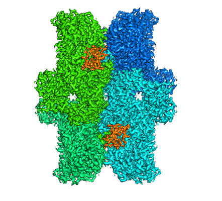
 Downloads & links
 emd_30650.png
 http://ftp.pdbj.org/pub/emdb/structures/EMD-30650


 Z
Z Y
Y X
X


 Sample components
 Processing
 Electron microscopy
 FIELD EMISSION GUN