National Natural Science Foundation of China (NSFC)
91940302, 91540201, 31430024, 31325007
China
Chinese Academy of Sciences
XDB37010201
China
Citation
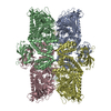
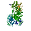
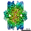
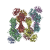
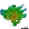
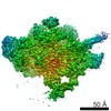
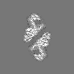
Journal: FEBS Open Bio / Year: 2020 Title: Cryo-EM structure of fission yeast tetrameric α-mannosidase Ams1. Authors: Jianxiu Zhang / Ying-Ying Wang / Li-Lin Du / Keqiong Ye / Abstract: Fungal α-mannosidase Ams1 and its mammalian homolog MAN2C1 hydrolyze terminal α-linked mannoses in free oligosaccharides released from misfolded glycoproteins or lipid-linked oligosaccharide donors. ...Fungal α-mannosidase Ams1 and its mammalian homolog MAN2C1 hydrolyze terminal α-linked mannoses in free oligosaccharides released from misfolded glycoproteins or lipid-linked oligosaccharide donors. Ams1 is transported by selective autophagy into vacuoles. Here, we determine the tetrameric structure of Ams1 from the fission yeast Schizosaccharomyces pombe at 3.2 Å resolution by cryo-electron microscopy. Distinct from a low resolution structure of S. cerevisiae Ams1, S. pombe Ams1 has a prominent N-terminal tail that mediates tetramerization and an extra β-sheet domain. Ams1 shares a conserved active site with other enzymes in glycoside hydrolase family 38, to which Ams1 belongs, but contains extra N-terminal domains involved in tetramerization. The atomic structure of Ams1 reported here will aid understanding of its enzymatic activity and transport mechanism.
History
Deposition
Feb 17, 2020
-
Header (metadata) release
Sep 9, 2020
-
Map release
Sep 9, 2020
-
Update
Mar 27, 2024
-
Current status
Mar 27, 2024
Processing site: PDBj / Status: Released
-
Structure visualization
Movie
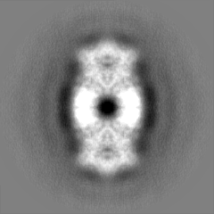
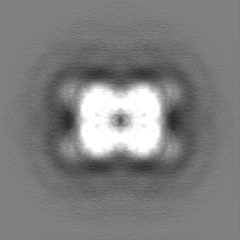
Surface view with section colored by density value
Name: ZINC ION / type: ligand / ID: 2 / Number of copies: 4 / Formula: ZN
Molecular weight
Theoretical: 65.409 Da
-
Experimental details
-
Structure determination
Method
cryo EM
Processing
single particle reconstruction
Aggregation state
particle
-
Sample preparation
Concentration
0.5 mg/mL
Buffer
pH: 7.5 Component:
Concentration
Formula
Name
50.0 mM
Tris-HCl
Tris hydrochloride
150.0 mM
NaCl
Sodium chloride
5.0 mM
MgCl2
Magnesium chloride
Grid
Model: Quantifoil R1.2/1.3 / Material: COPPER / Mesh: 300 / Support film - Material: CARBON / Support film - topology: HOLEY ARRAY / Pretreatment - Type: PLASMA CLEANING / Pretreatment - Time: 60 sec.
Vitrification
Cryogen name: ETHANE / Chamber humidity: 100 % / Chamber temperature: 277 K / Instrument: FEI VITROBOT MARK IV / Details: blot for 5 seconds before plunging.
-
Electron microscopy
Microscope
FEI TITAN KRIOS
Specialist optics
Phase plate: VOLTA PHASE PLATE / Energy filter - Name: GIF Tridiem 4K / Energy filter - Slit width: 20 eV
Image recording
Film or detector model: GATAN K2 SUMMIT (4k x 4k) / Detector mode: SUPER-RESOLUTION / Number real images: 597 / Average exposure time: 8.4 sec. / Average electron dose: 60.0 e/Å2
Electron beam
Acceleration voltage: 300 kV / Electron source: FIELD EMISSION GUN
In the structure databanks used in Yorodumi, some data are registered as the other names, "COVID-19 virus" and "2019-nCoV". Here are the details of the virus and the list of structure data.
Jan 31, 2019. EMDB accession codes are about to change! (news from PDBe EMDB page)
EMDB accession codes are about to change! (news from PDBe EMDB page)
The allocation of 4 digits for EMDB accession codes will soon come to an end. Whilst these codes will remain in use, new EMDB accession codes will include an additional digit and will expand incrementally as the available range of codes is exhausted. The current 4-digit format prefixed with “EMD-” (i.e. EMD-XXXX) will advance to a 5-digit format (i.e. EMD-XXXXX), and so on. It is currently estimated that the 4-digit codes will be depleted around Spring 2019, at which point the 5-digit format will come into force.
The EM Navigator/Yorodumi systems omit the EMD- prefix.
Related info.:Q: What is EMD? / ID/Accession-code notation in Yorodumi/EM Navigator
Yorodumi is a browser for structure data from EMDB, PDB, SASBDB, etc.
This page is also the successor to EM Navigator detail page, and also detail information page/front-end page for Omokage search.
The word "yorodu" (or yorozu) is an old Japanese word meaning "ten thousand". "mi" (miru) is to see.
Related info.:EMDB / PDB / SASBDB / Comparison of 3 databanks / Yorodumi Search / Aug 31, 2016. New EM Navigator & Yorodumi / Yorodumi Papers / Jmol/JSmol / Function and homology information / Changes in new EM Navigator and Yorodumi
 Movie
Movie Controller


 Open data
 Basic information
 Map data
 Sample
 Keywords
 Function and homology information
 Authors
 China, 3 items
  Citation
 Structure visualization
 Downloads & links
 emd_30021.png
 http://ftp.pdbj.org/pub/emdb/structures/EMD-30021
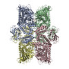


 Z (Sec.)
 Y (Row.)
 X (Col.)


 Sample components
 Processing
 Electron microscopy
 FIELD EMISSION GUN
