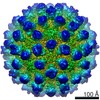
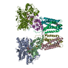
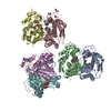
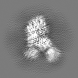
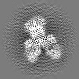
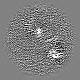
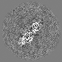
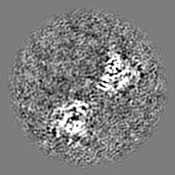
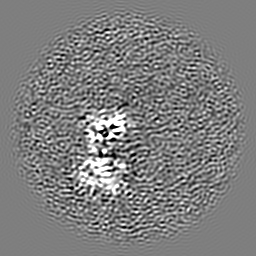
Mouse Norovirus Protruding domain complexed with neutralizing Fab fragment from mAb A6.2
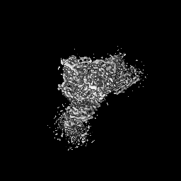
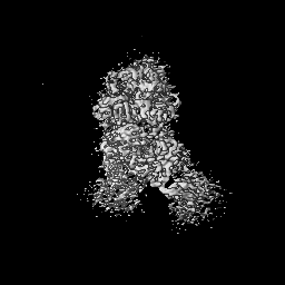
Map data
Soluble form of mouse norovirus protruding domain complexed with Fab fragments from neutralizing mAb A6.2
Sample
Complex: Murine norovirus 1 - Fab complex
Complex: Capsid protein
Protein or peptide: Capsid protein
Complex: Anti mouse norovirus mAb A6.2 Fab
Protein or peptide: Anti mouse norovirus mAb A6.2 Fab light chain
Protein or peptide: Anti mouse norovirus mAb A6.2 Fab heavy chain
Keywords
Antibody / norovirus / spike / VIRAL PROTEIN / VIRAL PROTEIN-IMMUNE SYSTEM complex
Function / homology
Function and homology information
antigenic variation / symbiont-mediated perturbation of host defense response / virion component / host cell cytoplasm Similarity search - Function
Calicivirus coat protein C-terminal / Calicivirus coat protein C-terminal / Calicivirus coat protein / Calicivirus coat protein / Picornavirus/Calicivirus coat protein / Viral coat protein subunit Similarity search - Domain/homology
National Institutes of Health/National Institute Of Allergy and Infectious Diseases (NIH/NIAID)
1R01-AI141465
United States
Citation
Journal: J Virol / Year: 2021 Title: A Norovirus Uses Bile Salts To Escape Antibody Recognition While Enhancing Receptor Binding. Authors: Alexis N Williams / Michael B Sherman / Hong Q Smith / Stefan Taube / B Montgomery Pettitt / Christiane E Wobus / Thomas J Smith / Abstract: Noroviruses, members of the family, are the major cause of epidemic gastroenteritis in humans, causing ∼20 million cases annually. These plus-strand RNA viruses have T=3 icosahedral protein ...Noroviruses, members of the family, are the major cause of epidemic gastroenteritis in humans, causing ∼20 million cases annually. These plus-strand RNA viruses have T=3 icosahedral protein capsids with 90 pronounced protruding (P) domain dimers to which antibodies and cellular receptors bind. In the case of mouse norovirus (MNV), bile salts have been shown to enhance receptor (CD300lf) binding to the P domain. We demonstrated previously that the P domains of several genotypes are markedly flexible and "float" over the shell, but the role of this flexibility was unclear. Recently, we demonstrated that bile causes a 90° rotation and collapse of the P domain onto the shell surface. Since bile binds distally to the P-shell interface, it was not at all clear how it could cause such dramatic changes. Here, we present the near-atomic resolution cryo-electron microscopy (cryo-EM) structure of the MNV protruding domain complexed with a neutralizing Fab. On the basis of previous results, we show here that bile salts cause allosteric conformational changes in the P domain that block antibody recognition of the top of the P domain. In addition, bile causes a major rearrangement of the P domain dimers that is likely responsible for the bile-induced collapse of the P domain onto the shell. In the contracted shell conformation, antibodies to the P1 and shell domains are not expected to bind. Therefore, at the site of infection in the gut, the host's own bile allows the virus to escape antibody-mediated neutralization while enhancing cell attachment. The major feature of calicivirus capsids is the 90 protruding domains (P domains) that are the site of cell receptor attachment and antibody epitopes. We demonstrated previously that these P domains are highly mobile and that bile causes these "floating" P domains in mouse norovirus (MNV) to contract onto the shell surface. Here, we present the near-atomic cryo-EM structure of the isolated MNV P domain complexed with a neutralizing Fab fragment. Our data show that bile causes two sets of changes. First, bile causes allosteric conformational changes in the epitopes at the top of the P domain that block antibody binding. Second, bile causes the P domain dimer subunits to rotate relative to each other, causing a contraction of the P domain that buries epitopes at the base of the P and shell domains. Taken together, the results show that MNV uses the host's own metabolites to enhance cell receptor binding while simultaneously blocking antibody recognition.
History
Deposition
Dec 22, 2020
-
Header (metadata) release
Apr 7, 2021
-
Map release
Apr 7, 2021
-
Update
Jun 4, 2025
-
Current status
Jun 4, 2025
Processing site: RCSB / Status: Released
-
Structure visualization
Movie
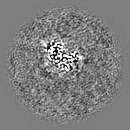
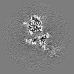
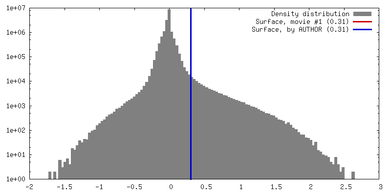
Surface view with section colored by density value
In the structure databanks used in Yorodumi, some data are registered as the other names, "COVID-19 virus" and "2019-nCoV". Here are the details of the virus and the list of structure data.
Jan 31, 2019. EMDB accession codes are about to change! (news from PDBe EMDB page)
EMDB accession codes are about to change! (news from PDBe EMDB page)
The allocation of 4 digits for EMDB accession codes will soon come to an end. Whilst these codes will remain in use, new EMDB accession codes will include an additional digit and will expand incrementally as the available range of codes is exhausted. The current 4-digit format prefixed with “EMD-” (i.e. EMD-XXXX) will advance to a 5-digit format (i.e. EMD-XXXXX), and so on. It is currently estimated that the 4-digit codes will be depleted around Spring 2019, at which point the 5-digit format will come into force.
The EM Navigator/Yorodumi systems omit the EMD- prefix.
Related info.:Q: What is EMD? / ID/Accession-code notation in Yorodumi/EM Navigator
Yorodumi is a browser for structure data from EMDB, PDB, SASBDB, etc.
This page is also the successor to EM Navigator detail page, and also detail information page/front-end page for Omokage search.
The word "yorodu" (or yorozu) is an old Japanese word meaning "ten thousand". "mi" (miru) is to see.
Related info.:EMDB / PDB / SASBDB / Comparison of 3 databanks / Yorodumi Search / Aug 31, 2016. New EM Navigator & Yorodumi / Yorodumi Papers / Jmol/JSmol / Function and homology information / Changes in new EM Navigator and Yorodumi
 Movie
Movie Controller

 Yorodumi
 Open data
 Basic information
 Map data
 Sample
 Keywords
 Function and homology information
 Murine norovirus 1 /
 Authors
 United States, 1 items
  Citation
 Structure visualization
 Downloads & links
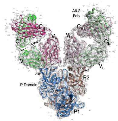 emd_23187.png
 http://ftp.pdbj.org/pub/emdb/structures/EMD-23187


 Z (Sec.)
 Y (Row.)
 X (Col.)


 Sample components
 Processing
 Electron microscopy
 FIELD EMISSION GUN
