Movie
Movie Controller


+ Open data

- Basic information
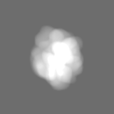
| Entry | Database: EMDB / ID: EMD-22422 | |||||||||
|---|---|---|---|---|---|---|---|---|---|---|
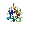
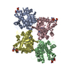
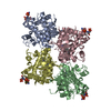
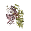
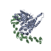
| Title | Structure of an endocytic receptor | |||||||||
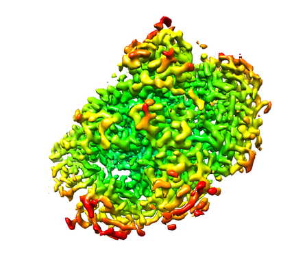
 Map data Map data | Final map | |||||||||
 Sample Sample |
| |||||||||
 Keywords Keywords | Cell-surface receptor / Immune receptor / mannose receptor family / IMMUNE SYSTEM | |||||||||
| Function / homology |  Function and homology information Function and homology informationendocytosis / carbohydrate binding / signaling receptor activity / immune response / inflammatory response / external side of plasma membrane / extracellular exosome / plasma membrane Similarity search - Function | |||||||||
| Biological species |  Homo sapiens (human) Homo sapiens (human) | |||||||||
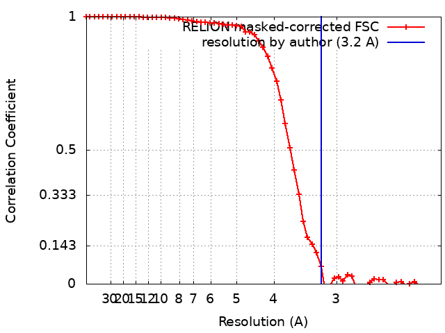
| Method | single particle reconstruction / cryo EM / Resolution: 3.2 Å | |||||||||
 Authors Authors | Gully BS / Rossjohn J | |||||||||
| Funding support |  Australia, 2 items Australia, 2 items
| |||||||||
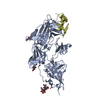
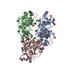
 Citation Citation | Journal: J Biol Chem / Year: 2021 Title: The cryo-EM structure of the endocytic receptor DEC-205. Authors: Benjamin S Gully / Hariprasad Venugopal / Alex J Fulcher / Zhihui Fu / Jessica Li / Felix A Deuss / Carmen Llerena / William R Heath / Mireille H Lahoud / Irina Caminschi / Jamie Rossjohn / Richard Berry /  Abstract: DEC-205 (CD205), a member of the macrophage mannose receptor protein family, is the prototypic endocytic receptor of dendritic cells, whose ligands include phosphorothioated cytosine-guanosine ...DEC-205 (CD205), a member of the macrophage mannose receptor protein family, is the prototypic endocytic receptor of dendritic cells, whose ligands include phosphorothioated cytosine-guanosine oligonucleotides, a motif often seen in bacterial or viral DNA. However, despite growing biological and clinical significance, little is known about the structural arrangement of this receptor or any of its family members. Here, we describe the 3.2 Å cryo-EM structure of human DEC-205, thereby illuminating the structure of the mannose receptor protein family. The DEC-205 monomer forms a compact structure comprising two intercalated rings of C-type lectin-like domains, where the N-terminal cysteine-rich and fibronectin domains reside at the central intersection. We establish a pH-dependent oligomerization pathway forming tetrameric DEC-205 using solution-based techniques and ultimately solved the 4.9 Å cryo-EM structure of the DEC-205 tetramer to identify the unfurling of the second lectin ring which enables tetramer formation. Furthermore, we suggest the relevance of this oligomerization pathway within a cellular setting, whereby cytosine-guanosine binding appeared to disrupt this cell-surface oligomer. Accordingly, we provide insight into the structure and oligomeric assembly of the DEC-205 receptor. | |||||||||
| History |
|
- Structure visualization
| Movie |
Movie viewer |
|---|---|
| Structure viewer | EM map: SurfViewMolmilJmol/JSmol |
| Supplemental images |
- Downloads & links
-EMDB archive
| Map data | emd_22422.map.gz | 2.4 MB | EMDB map data format | |
|---|---|---|---|---|
| Header (meta data) | emd-22422-v30.xmlemd-22422.xml | 19.3 KB 19.3 KB | Display Display | EMDB header |
| FSC (resolution estimation) | emd_22422_fsc.xml | 5.8 KB | Display | FSC data file |
| Images |  emd_22422.png emd_22422.png | 151.6 KB | ||
| Masks | emd_22422_msk_1.map | 15.6 MB | Mask map | |
| Filedesc metadata | emd-22422.cif.gz | 7 KB | ||
| Others | emd_22422_half_map_1.map.gzemd_22422_half_map_2.map.gz | 11.9 MB 11.9 MB | ||
| Archive directory |  http://ftp.pdbj.org/pub/emdb/structures/EMD-22422ftp://ftp.pdbj.org/pub/emdb/structures/EMD-22422 http://ftp.pdbj.org/pub/emdb/structures/EMD-22422ftp://ftp.pdbj.org/pub/emdb/structures/EMD-22422 | HTTPS FTP |
-Related structure data
| Related structure data |  7jptMC  7jpuC M: atomic model generated by this map C: citing same article ( |
|---|---|
| Similar structure data |


-Links
| EMDB pages | EMDB (EBI/PDBe) / EMDataResource |
|---|---|
| Related items in Molecule of the Month |


-Map
| File | Download / File: emd_22422.map.gz / Format: CCP4 / Size: 15.6 MB / Type: IMAGE STORED AS FLOATING POINT NUMBER (4 BYTES) | ||||||||||||||||||||||||||||||||||||||||||||||||||||||||||||||||||||
|---|---|---|---|---|---|---|---|---|---|---|---|---|---|---|---|---|---|---|---|---|---|---|---|---|---|---|---|---|---|---|---|---|---|---|---|---|---|---|---|---|---|---|---|---|---|---|---|---|---|---|---|---|---|---|---|---|---|---|---|---|---|---|---|---|---|---|---|---|---|
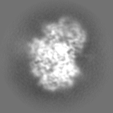
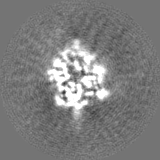
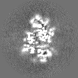
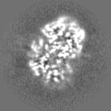
| Annotation | Final map | ||||||||||||||||||||||||||||||||||||||||||||||||||||||||||||||||||||
| Projections & slices | Image control
Images are generated by Spider. | ||||||||||||||||||||||||||||||||||||||||||||||||||||||||||||||||||||
| Voxel size | X=Y=Z: 1.06 Å | ||||||||||||||||||||||||||||||||||||||||||||||||||||||||||||||||||||
| Density |
| ||||||||||||||||||||||||||||||||||||||||||||||||||||||||||||||||||||
| Symmetry | Space group: 1 | ||||||||||||||||||||||||||||||||||||||||||||||||||||||||||||||||||||
| Details | EMDB XML:
CCP4 map header:
| ||||||||||||||||||||||||||||||||||||||||||||||||||||||||||||||||||||
 Z (Sec.)
 Y (Row.)
 X (Col.)


-Supplemental data
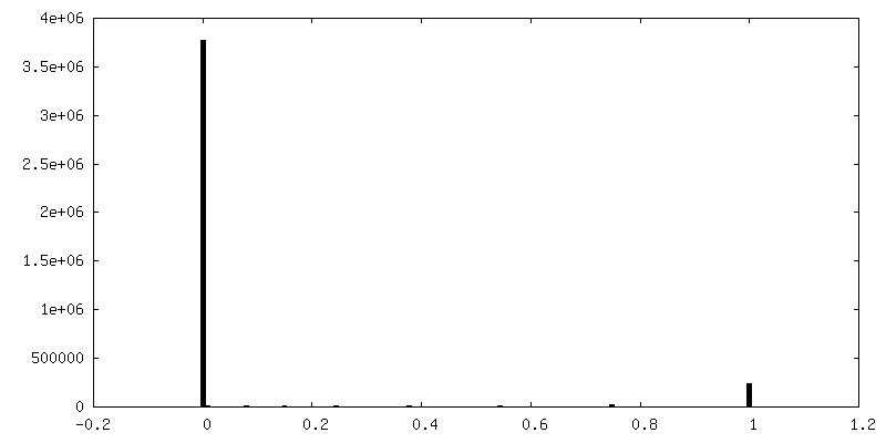
-Mask #1
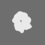
| File | emd_22422_msk_1.map | ||||||||||||
|---|---|---|---|---|---|---|---|---|---|---|---|---|---|
| Projections & Slices |
| ||||||||||||
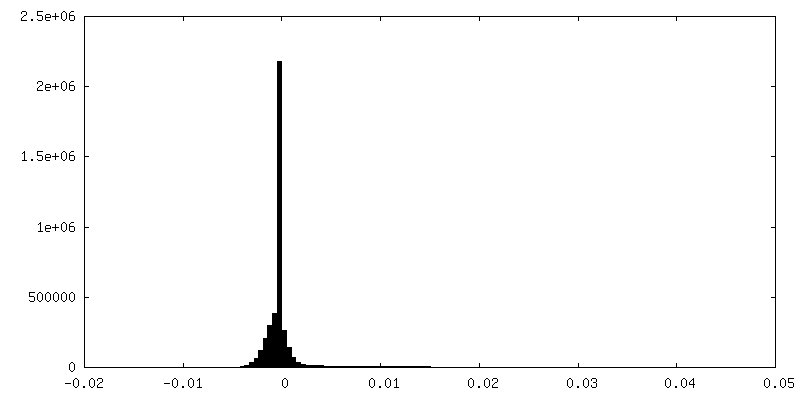
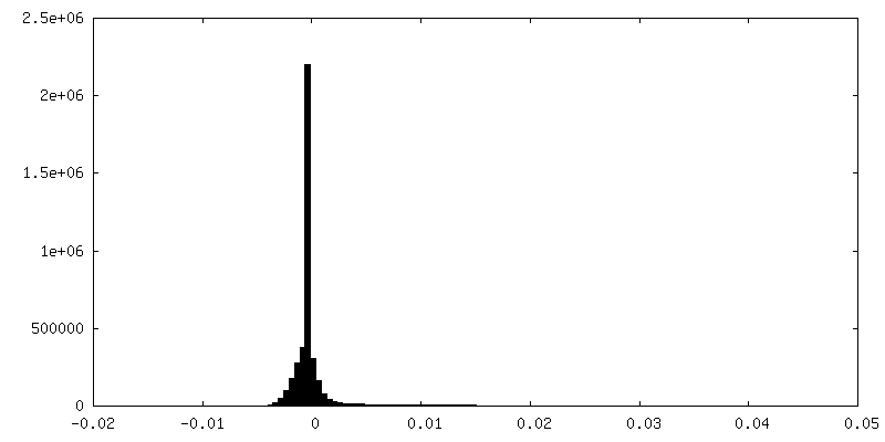
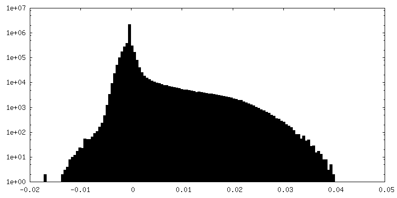
| Density Histograms |

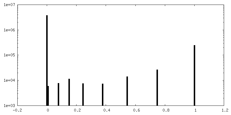
-Half map: half map 1
| File | emd_22422_half_map_1.map | ||||||||||||
|---|---|---|---|---|---|---|---|---|---|---|---|---|---|
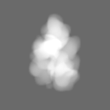
| Annotation | half map 1 | ||||||||||||
| Projections & Slices |
| ||||||||||||
| Density Histograms |


-Half map: half map 2
| File | emd_22422_half_map_2.map | ||||||||||||
|---|---|---|---|---|---|---|---|---|---|---|---|---|---|
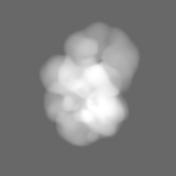
| Annotation | half map 2 | ||||||||||||
| Projections & Slices |
| ||||||||||||
| Density Histograms |


- Sample components
-Entire : Lymphocyte antigen 75, DEC205, CD205
| Entire | Name: Lymphocyte antigen 75, DEC205, CD205 |
|---|---|
| Components |
|
-Supramolecule #1: Lymphocyte antigen 75, DEC205, CD205
| Supramolecule | Name: Lymphocyte antigen 75, DEC205, CD205 / type: organelle_or_cellular_component / ID: 1 / Parent: 0 / Macromolecule list: #1 |
|---|---|
| Source (natural) | Organism: Homo sapiens (human) |
| Molecular weight | Theoretical: 200 KDa |
-Macromolecule #1: Lymphocyte antigen 75
| Macromolecule | Name: Lymphocyte antigen 75 / type: protein_or_peptide / ID: 1 / Number of copies: 1 / Enantiomer: LEVO |
|---|---|
| Source (natural) | Organism: Homo sapiens (human) |
| Molecular weight | Theoretical: 195.23025 KDa |
| Recombinant expression | Organism: Homo sapiens (human) |
| Sequence | String: RAANDPFTIV HGNTGKCIKP VYGWIVADDC DETEDKLWKW VSQHRLFHLH SQKCLGLDIT KSVNELRMFS CDSSAMLWWK CEHHSLYGA ARYRLALKDG HGTAISNASD VWKKGGSEES LCDQPYHEIY TRDGNSYGRP CEFPFLIDGT WHHDCILDED H SGPWCATT ...String: RAANDPFTIV HGNTGKCIKP VYGWIVADDC DETEDKLWKW VSQHRLFHLH SQKCLGLDIT KSVNELRMFS CDSSAMLWWK CEHHSLYGA ARYRLALKDG HGTAISNASD VWKKGGSEES LCDQPYHEIY TRDGNSYGRP CEFPFLIDGT WHHDCILDED H SGPWCATT LNYEYDRKWG ICLKPENGCE DNWEKNEQFG SCYQFNTQTA LSWKEAYVSC QNQGADLLSI NSAAELTYLK EK EGIAKIF WIGLNQLYSA RGWEWSDHKP LNFLNWDPDR PSAPTIGGSS CARMDAESGL WQSFSCEAQL PYVCRKPLNN TVE LTDVWT YSDTRCDAGW LPNNGFCYLL VNESNSWDKA HAKCKAFSSD LISIHSLADV EVVVTKLHNE DIKEEVWIGL KNIN IPTLF QWSDGTEVTL TYWDENEPNV PYNKTPNCVS YLGELGQWKV QSCEEKLKYV CKRKGEKLND ASSDKMCPPD EGWKR HGET CYKIYEDEVP FGTNCNLTIT SRFEQEYLND LMKKYDKSLR KYFWTGLRDV DSCGEYNWAT VGGRRRAVTF SNWNFL EPA SPGGCVAMST GKSVGKWEVK DCRSFKALSI CKKMSGPLGP EEASPKPDDP CPEGWQSFPA SLSCYKVFHA ERIVRKR NW EEAERFCQAL GAHLSSFSHV DEIKEFLHFL TDQFSGQHWL WIGLNKRSPD LQGSWQWSDR TPVSTIIMPN EFQQDYDI R DCAAVKVFHR PWRRGWHFYD DREFIYLRPF ACDTKLEWVC QIPKGRTPKT PDWYNPDRAG IHGPPLIIEG SEYWFVADL HLNYEEAVLY CASNHSFLAT ITSFVGLKAI KNKIANISGD GQKWWIRISE WPIDDHFTYS RYPWHRFPVT FGEECLYMSA KTWLIDLGK PTDCSTKLPF ICEKYNVSSL EKYSPDSAAK VQCSEQWIPF QNKCFLKIKP VSLTFSQASD TCHSYGGTLP S VLSQIEQD FITSLLPDME ATLWIGLRWT AYEKINKWTD NRELTYSNFH PLLVSGRLRI PENFFEEESR YHCALILNLQ KS PFTGTWN FTSCSERHFV SLCQKYSEVK SRQTLQNASE TVKYLNNLYK IIPKTLTWHS AKRECLKSNM QLVSITDPYQ QAF LSVQAL LHNSSLWIGL FSQDDELNFG WSDGKRLHFS RWAETNGQLE DCVVLDTDGF WKTVDCNDNQ PGAICYYSGN ETEK EVKPV DSVKCPSPVL NTPWIPFQNC CYNFIITKNR HMATTQDEVH TKCQKLNPKS HILSIRDEKE NNFVLEQLLY FNYMA SWVM LGITYRNKSL MWFDKTPLSY THWRAGRPTI KNEKFLAGLS TDGFWDIQTF KVIEEAVYFH QHSILACKIE MVDYKE EYN TTLPQFMPYE DGIYSVIQKK VTWYEALNMC SQSGGHLASV HNQNGQLFLE DIVKRDGFPL WVGLSSHDGS ESSFEWS DG STFDYIPWKG QTSPGNCVLL DPKGTWKHEK CNSVKDGAIC YKPTKSKKLS RLTYSSRCPA AKENGSRWIQ YKGHCYKS D QALHSFSEAK KLCSKHDHSA TIVSIKDEDE NKFVSRLMRE NNNITMRVWL GLSQHSVDQS WSWLDGSEVT FVKWENKSK SGVGRCSMLI ASNETWKKVE CEHGFGRVVC KVPLGPDYTA IAIIVATLSI LVLMGGLIWF LFQRHRLHLA GFSSVRYAQG VNEDEIMLP SFHD UniProtKB: Lymphocyte antigen 75 |
-Macromolecule #3: 2-acetamido-2-deoxy-beta-D-glucopyranose
| Macromolecule | Name: 2-acetamido-2-deoxy-beta-D-glucopyranose / type: ligand / ID: 3 / Number of copies: 2 / Formula: NAG |
|---|---|
| Molecular weight | Theoretical: 221.208 Da |
| Chemical component information |  ChemComp-NAG: |
-Experimental details
-Structure determination
| Method | cryo EM |
|---|---|
 Processing Processing | single particle reconstruction |
| Aggregation state | particle |
-Sample preparation
| Concentration | 0.1 mg/mL | ||||||
|---|---|---|---|---|---|---|---|
| Buffer | pH: 8 Component:
Details: 20 mM Tris-HCl at pH 8.0, 150 mM NaCl buffer | ||||||
| Vitrification | Cryogen name: ETHANE / Chamber humidity: 100 % / Instrument: FEI VITROBOT MARK II | ||||||
| Details | 3 microlitres of sample in 20 mM Tris-HCl at pH 8.0, 150 mM NaCl buffer |
- Electron microscopy
| Microscope | FEI TITAN KRIOS |
|---|---|
| Image recording | Film or detector model: GATAN K2 SUMMIT (4k x 4k) / Detector mode: COUNTING / Average electron dose: 63.0 e/Å2 |
| Electron beam | Acceleration voltage: 300 kV / Electron source:  FIELD EMISSION GUN FIELD EMISSION GUN |
| Electron optics | Illumination mode: FLOOD BEAM / Imaging mode: BRIGHT FIELD |
| Sample stage | Specimen holder model: FEI TITAN KRIOS AUTOGRID HOLDER / Cooling holder cryogen: NITROGEN |
| Experimental equipment |  Model: Titan Krios / Image courtesy: FEI Company |
+Image processing
-Atomic model buiding 1
| Initial model |
| ||||||||
|---|---|---|---|---|---|---|---|---|---|
| Refinement | Space: REAL / Protocol: RIGID BODY FIT | ||||||||
| Output model | PDB-7jpt: |