Lon AAA+ / protease / cryo-EM / dual pore-loops / MOTOR PROTEIN
Function / homology
Function and homology information
endopeptidase La / ATP-dependent peptidase activity / protein quality control for misfolded or incompletely synthesized proteins / cellular response to heat / sequence-specific DNA binding / serine-type endopeptidase activity / ATP hydrolysis activity / ATP binding / metal ion binding / identical protein binding / cytoplasm Similarity search - Function
Lon protease, bacterial / : / Lon protease, bacterial/eukaryotic-type / Lon protease AAA+ ATPase lid domain / Peptidase S16, active site / ATP-dependent serine proteases, lon family, serine active site. / Lon proteolytic domain profile. / Peptidase S16, Lon proteolytic domain / Lon protease / Lon protease (S16) C-terminal proteolytic domain ...Lon protease, bacterial / : / Lon protease, bacterial/eukaryotic-type / Lon protease AAA+ ATPase lid domain / Peptidase S16, active site / ATP-dependent serine proteases, lon family, serine active site. / Lon proteolytic domain profile. / Peptidase S16, Lon proteolytic domain / Lon protease / Lon protease (S16) C-terminal proteolytic domain / Lon N-terminal domain profile. / Lon protease, N-terminal domain / Lon protease, N-terminal domain superfamily / ATP-dependent protease La (LON) substrate-binding domain / Found in ATP-dependent protease La (LON) / PUA-like superfamily / ATPase family associated with various cellular activities (AAA) / ATPase, AAA-type, core / Ribosomal protein S5 domain 2-type fold, subgroup / Ribosomal protein S5 domain 2-type fold / ATPases associated with a variety of cellular activities / AAA+ ATPase domain / P-loop containing nucleoside triphosphate hydrolase Similarity search - Domain/homology
National Institutes of Health/National Institute of General Medical Sciences (NIH/NIGMS)
P41GM103832
United States
National Institutes of Health/National Institute of General Medical Sciences (NIH/NIGMS)
R01GM079429
United States
National Institutes of Health/National Institute of General Medical Sciences (NIH/NIGMS)
S10OD021600
United States
Citation
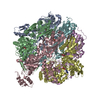
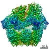
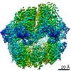
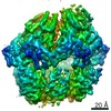
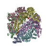
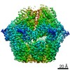
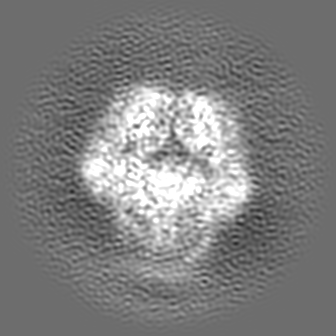
Journal: J Biol Chem / Year: 2021 Title: Molecular basis for ATPase-powered substrate translocation by the Lon AAA+ protease. Authors: Shanshan Li / Kan-Yen Hsieh / Shih-Chieh Su / Grigore D Pintilie / Kaiming Zhang / Chung-I Chang / Abstract: The Lon AAA+ (adenosine triphosphatases associated with diverse cellular activities) protease (LonA) converts ATP-fuelled conformational changes into sufficient mechanical force to drive ...The Lon AAA+ (adenosine triphosphatases associated with diverse cellular activities) protease (LonA) converts ATP-fuelled conformational changes into sufficient mechanical force to drive translocation of a substrate into a hexameric proteolytic chamber. To understand the structural basis for the substrate translocation process, we determined the cryo-electron microscopy (cryo-EM) structure of Meiothermus taiwanensis LonA (MtaLonA) in a substrate-engaged state at 3.6 Å resolution. Our data indicate that substrate interactions are mediated by the dual pore loops of the ATPase domains, organized in spiral staircase arrangement from four consecutive protomers in different ATP-binding and hydrolysis states. However, a closed AAA+ ring is maintained by two disengaged ADP-bound protomers transiting between the lowest and highest position. This structure reveals a processive rotary translocation mechanism mediated by LonA-specific nucleotide-dependent allosteric coordination among the ATPase domains, which is induced by substrate binding.
History
Deposition
Apr 28, 2020
-
Header (metadata) release
Jun 9, 2021
-
Map release
Jun 9, 2021
-
Update
Oct 16, 2024
-
Current status
Oct 16, 2024
Processing site: RCSB / Status: Released
-
Structure visualization
Movie
Surface view with section colored by density value
Cryogen name: ETHANE / Chamber humidity: 100 % / Instrument: FEI VITROBOT MARK IV
-
Electron microscopy
Microscope
FEI TITAN KRIOS
Image recording
Film or detector model: GATAN K2 SUMMIT (4k x 4k) / Detector mode: COUNTING / Number real images: 1800 / Average exposure time: 6.0 sec. / Average electron dose: 8.5 e/Å2
Electron beam
Acceleration voltage: 300 kV / Electron source: FIELD EMISSION GUN
Electron optics
C2 aperture diameter: 70.0 µm / Illumination mode: FLOOD BEAM / Imaging mode: BRIGHT FIELD / Cs: 2.7 mm
In the structure databanks used in Yorodumi, some data are registered as the other names, "COVID-19 virus" and "2019-nCoV". Here are the details of the virus and the list of structure data.
Jan 31, 2019. EMDB accession codes are about to change! (news from PDBe EMDB page)
EMDB accession codes are about to change! (news from PDBe EMDB page)
The allocation of 4 digits for EMDB accession codes will soon come to an end. Whilst these codes will remain in use, new EMDB accession codes will include an additional digit and will expand incrementally as the available range of codes is exhausted. The current 4-digit format prefixed with “EMD-” (i.e. EMD-XXXX) will advance to a 5-digit format (i.e. EMD-XXXXX), and so on. It is currently estimated that the 4-digit codes will be depleted around Spring 2019, at which point the 5-digit format will come into force.
The EM Navigator/Yorodumi systems omit the EMD- prefix.
Related info.:Q: What is EMD? / ID/Accession-code notation in Yorodumi/EM Navigator
Yorodumi is a browser for structure data from EMDB, PDB, SASBDB, etc.
This page is also the successor to EM Navigator detail page, and also detail information page/front-end page for Omokage search.
The word "yorodu" (or yorozu) is an old Japanese word meaning "ten thousand". "mi" (miru) is to see.
Related info.:EMDB / PDB / SASBDB / Comparison of 3 databanks / Yorodumi Search / Aug 31, 2016. New EM Navigator & Yorodumi / Yorodumi Papers / Jmol/JSmol / Function and homology information / Changes in new EM Navigator and Yorodumi
 Movie
Movie Controller

 Yorodumi
 Open data
 Basic information
 Map data
 Sample
 Keywords
 Function and homology information
 Meiothermus taiwanensis (bacteria) /
 Authors
 United States, 3 items
  Citation

 Structure visualization
 Downloads & links
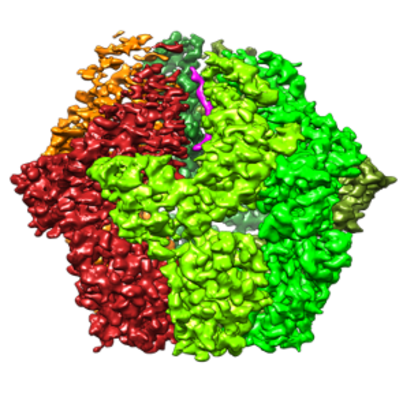 emd_21870.png
 http://ftp.pdbj.org/pub/emdb/structures/EMD-21870
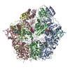


 Z (Sec.)
 Y (Row.)
 X (Col.)


 Sample components

 Processing
 Electron microscopy
 FIELD EMISSION GUN