Movie
Movie Controller

[English] 日本語
 Yorodumi
- EMDB-16780: CryoEM structure of AL55 amyloid fibrils extracted from the kidne... -
+ Open data

- Basic information
| Entry |  | |||||||||
|---|---|---|---|---|---|---|---|---|---|---|
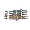
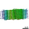
| Title | CryoEM structure of AL55 amyloid fibrils extracted from the kidney of an AL amyloidosis patient. | |||||||||
 Map data Map data | ||||||||||
 Sample Sample |
| |||||||||
 Keywords Keywords | Light chains / Amyloid fibrils / AL amyloidosis. / PROTEIN FIBRIL | |||||||||
| Biological species |  Homo sapiens (human) Homo sapiens (human) | |||||||||
| Method | helical reconstruction / cryo EM / Resolution: 4.0 Å | |||||||||
 Authors Authors | Puri S / Schulte T / Chaves-Sanjuan A / Ricagno S | |||||||||
| Funding support |  Italy, 1 items Italy, 1 items
| |||||||||
 Citation Citation | Journal: J Mol Biol / Year: 2023 Title: The Cryo-EM STRUCTURE of Renal Amyloid Fibril Suggests Structurally Homogeneous Multiorgan Aggregation in AL Amyloidosis. Authors: Sarita Puri / Tim Schulte / Antonio Chaves-Sanjuan / Giulia Mazzini / Serena Caminito / Carlo Pappone / Luigi Anastasia / Paolo Milani / Giampaolo Merlini / Martino Bolognesi / Mario ...Authors: Sarita Puri / Tim Schulte / Antonio Chaves-Sanjuan / Giulia Mazzini / Serena Caminito / Carlo Pappone / Luigi Anastasia / Paolo Milani / Giampaolo Merlini / Martino Bolognesi / Mario Nuvolone / Giovanni Palladini / Stefano Ricagno / Abstract: Immunoglobulin light chain amyloidosis (AL) is caused by the aberrant production of amyloidogenic light chains (LC) that accumulate as amyloid deposits in vital organs. Distinct LC sequences in each ...Immunoglobulin light chain amyloidosis (AL) is caused by the aberrant production of amyloidogenic light chains (LC) that accumulate as amyloid deposits in vital organs. Distinct LC sequences in each patient yield distinct amyloid structures. However different tissue microenvironments may also cause identical protein precursors to adopt distinct amyloid structures. To address the impact of the tissue environment on the structural polymorphism of amyloids, we extracted fibrils from the kidney of an AL patient (AL55) whose cardiac amyloid structure was previously determined by our group. Here we show that the 4.0 Å resolution cryo-EM structure of the renal fibril is virtually identical to that reported for the cardiac fibril. These results provide the first structural evidence that LC amyloids independently deposited in different organs of the same AL patient share a common fold. | |||||||||
| History |
|
- Structure visualization
| Supplemental images |
|---|

- Downloads & links
-EMDB archive
| Map data | emd_16780.map.gz | 2.8 MB |  EMDB map data format EMDB map data format | |
|---|---|---|---|---|
| Header (meta data) | emd-16780-v30.xmlemd-16780.xml | 15.5 KB 15.5 KB | Display Display | EMDB header |
| FSC (resolution estimation) | emd_16780_fsc.xml | 9.1 KB | Display | FSC data file |
| Images |  emd_16780.png emd_16780.png | 41.8 KB | ||
| Filedesc metadata | emd-16780.cif.gz | 5.4 KB | ||
| Others | emd_16780_half_map_1.map.gzemd_16780_half_map_2.map.gz | 49.3 MB 49.3 MB | ||
| Archive directory |  http://ftp.pdbj.org/pub/emdb/structures/EMD-16780ftp://ftp.pdbj.org/pub/emdb/structures/EMD-16780 http://ftp.pdbj.org/pub/emdb/structures/EMD-16780ftp://ftp.pdbj.org/pub/emdb/structures/EMD-16780 | HTTPS FTP |
-Related structure data
-Links
| EMDB pages | EMDB (EBI/PDBe) / EMDataResource |
|---|
-Map
| File | Download / File: emd_16780.map.gz / Format: CCP4 / Size: 64 MB / Type: IMAGE STORED AS FLOATING POINT NUMBER (4 BYTES) | ||||||||||||||||||||||||||||||||||||
|---|---|---|---|---|---|---|---|---|---|---|---|---|---|---|---|---|---|---|---|---|---|---|---|---|---|---|---|---|---|---|---|---|---|---|---|---|---|
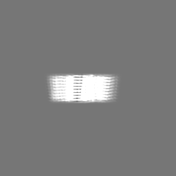
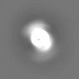
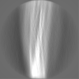
| Projections & slices | Image control
Images are generated by Spider. | ||||||||||||||||||||||||||||||||||||
| Voxel size | X=Y=Z: 1.11686 Å | ||||||||||||||||||||||||||||||||||||
| Density |
| ||||||||||||||||||||||||||||||||||||
| Symmetry | Space group: 1 | ||||||||||||||||||||||||||||||||||||
| Details | EMDB XML:
|
 Z (Sec.)
 Y (Row.)
 X (Col.)


-Supplemental data
-Half map: #2
| File | emd_16780_half_map_1.map | ||||||||||||
|---|---|---|---|---|---|---|---|---|---|---|---|---|---|
| Projections & Slices |
| ||||||||||||
| Density Histograms |


-Half map: #1
| File | emd_16780_half_map_2.map | ||||||||||||
|---|---|---|---|---|---|---|---|---|---|---|---|---|---|
| Projections & Slices |
| ||||||||||||
| Density Histograms |


- Sample components
-Entire : Renal AL55 amyloid fibrils
| Entire | Name: Renal AL55 amyloid fibrils |
|---|---|
| Components |
|
-Supramolecule #1: Renal AL55 amyloid fibrils
| Supramolecule | Name: Renal AL55 amyloid fibrils / type: complex / ID: 1 / Parent: 0 / Macromolecule list: all Details: Light chain amyloid fibrils named AL55 extracted from the kidney of an AL amyloidosis patient |
|---|---|
| Source (natural) | Organism: Homo sapiens (human) / Organ: Kidney |
-Macromolecule #1: Immunoglobulin lambda light chain
| Macromolecule | Name: Immunoglobulin lambda light chain / type: protein_or_peptide / ID: 1 / Number of copies: 5 / Enantiomer: LEVO |
|---|---|
| Source (natural) | Organism: Homo sapiens (human) / Organ: Kidney |
| Molecular weight | Theoretical: 23.326557 KDa |
| Sequence | String: NFMLTQPHSV SESPGKTLTI SCTGSSASIA SHYVQWYQQR PGGAPTTLIY ENDQRPSEVP DRFSGSIDSS SNSASLTISG LKTEDEADY YCQSYDGNNH WVFGGGTKLT VLSQPKAAPS VTLFPPSSEE LQANKATLVC LISDFYPGAV TVAWKADSSP V KAGVETTT ...String: NFMLTQPHSV SESPGKTLTI SCTGSSASIA SHYVQWYQQR PGGAPTTLIY ENDQRPSEVP DRFSGSIDSS SNSASLTISG LKTEDEADY YCQSYDGNNH WVFGGGTKLT VLSQPKAAPS VTLFPPSSEE LQANKATLVC LISDFYPGAV TVAWKADSSP V KAGVETTT PSKQSNNKYA ASSYLSLTPE QWKSHKSYSC QVTHEGSTVE KTVAPTECS |
-Experimental details
-Structure determination
| Method | cryo EM |
|---|---|
 Processing Processing | helical reconstruction |
| Aggregation state | filament |
-Sample preparation
| Buffer | pH: 7 |
|---|---|
| Vitrification | Cryogen name: ETHANE / Chamber humidity: 100 % / Chamber temperature: 277 K |
- Electron microscopy
| Microscope | FEI TALOS ARCTICA |
|---|---|
| Image recording | Film or detector model: FEI FALCON III (4k x 4k) / Number real images: 1819 / Average electron dose: 50.0 e/Å2 |
| Electron beam | Acceleration voltage: 200 kV / Electron source:  FIELD EMISSION GUN FIELD EMISSION GUN |
| Electron optics | Illumination mode: FLOOD BEAM / Imaging mode: BRIGHT FIELD / Nominal defocus max: 2.5 µm / Nominal defocus min: 0.8 µm |
| Sample stage | Specimen holder model: FEI TITAN KRIOS AUTOGRID HOLDER / Cooling holder cryogen: NITROGEN |
| Experimental equipment |  Model: Talos Arctica / Image courtesy: FEI Company |