Movie
Movie Controller

[English] 日本語
 Yorodumi
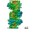
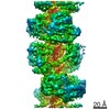
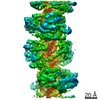
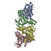
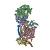
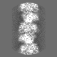
- EMDB-11937: CryoEM structure of the MDA5-dsRNA filament in complex with ADP w... -
+ Open data

- Basic information
| Entry | Database: EMDB / ID: EMD-11937 | |||||||||
|---|---|---|---|---|---|---|---|---|---|---|
| Title | CryoEM structure of the MDA5-dsRNA filament in complex with ADP with 92-degree helical twist | |||||||||
 Map data Map data | ||||||||||
 Sample Sample |
| |||||||||
| Function / homology |  Function and homology information Function and homology informationMDA-5 signaling pathway / positive regulation of response to cytokine stimulus / Ub-specific processing proteases / mRNA transcription / negative regulation of viral genome replication / type I interferon-mediated signaling pathway / cellular response to exogenous dsRNA / pattern recognition receptor activity / protein complex oligomerization / positive regulation of interferon-alpha production ...MDA-5 signaling pathway / positive regulation of response to cytokine stimulus / Ub-specific processing proteases / mRNA transcription / negative regulation of viral genome replication / type I interferon-mediated signaling pathway / cellular response to exogenous dsRNA / pattern recognition receptor activity / protein complex oligomerization / positive regulation of interferon-alpha production / protein sumoylation / ribonucleoprotein complex binding / positive regulation of interferon-beta production / antiviral innate immune response / cellular response to virus / positive regulation of interleukin-6 production / response to virus / positive regulation of tumor necrosis factor production / double-stranded RNA binding / defense response to virus / RNA helicase activity / single-stranded RNA binding / RNA helicase / protein domain specific binding / innate immune response / ATP hydrolysis activity / mitochondrion / DNA binding / RNA binding / zinc ion binding / ATP binding / identical protein binding / nucleus / cytoplasm Similarity search - Function | |||||||||
| Biological species |   Pseudomonas virus phi6 Pseudomonas virus phi6 | |||||||||
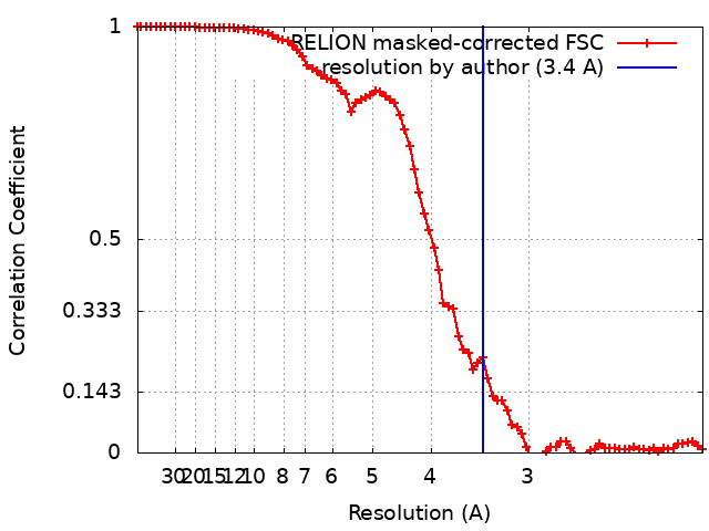
| Method | helical reconstruction / cryo EM / Resolution: 3.4 Å | |||||||||
 Authors Authors | Yu Q / Modis Y | |||||||||
| Funding support |  United Kingdom, 2 items United Kingdom, 2 items
| |||||||||
 Citation Citation | Journal: Nat Commun / Year: 2021 Title: MDA5 disease variant M854K prevents ATP-dependent structural discrimination of viral and cellular RNA. Authors: Qin Yu / Alba Herrero Del Valle / Rahul Singh / Yorgo Modis /  Abstract: Our innate immune responses to viral RNA are vital defenses. Long cytosolic double-stranded RNA (dsRNA) is recognized by MDA5. The ATPase activity of MDA5 contributes to its dsRNA binding selectivity. ...Our innate immune responses to viral RNA are vital defenses. Long cytosolic double-stranded RNA (dsRNA) is recognized by MDA5. The ATPase activity of MDA5 contributes to its dsRNA binding selectivity. Mutations that reduce RNA selectivity can cause autoinflammatory disease. Here, we show how the disease-associated MDA5 variant M854K perturbs MDA5-dsRNA recognition. M854K MDA5 constitutively activates interferon signaling in the absence of exogenous RNA. M854K MDA5 lacks ATPase activity and binds more stably to synthetic Alu:Alu dsRNA. CryoEM structures of MDA5-dsRNA filaments at different stages of ATP hydrolysis show that the K854 sidechain forms polar bonds that constrain the conformation of MDA5 subdomains, disrupting key steps in the ATPase cycle- RNA footprint expansion and helical twist modulation. The M854K mutation inhibits ATP-dependent RNA proofreading via an allosteric mechanism, allowing MDA5 to form signaling complexes on endogenous RNAs. This work provides insights on how MDA5 recognizes dsRNA in health and disease. | |||||||||
| History |
|
- Structure visualization
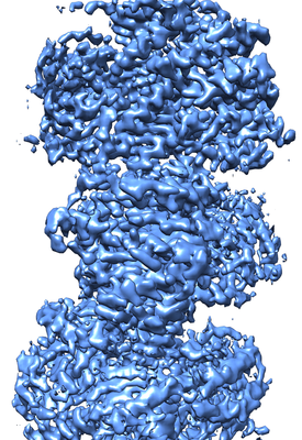
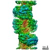
| Movie |
Movie viewer |
|---|---|
| Structure viewer | EM map: SurfViewMolmilJmol/JSmol |
| Supplemental images |
- Downloads & links
-EMDB archive
| Map data | emd_11937.map.gz | 6.2 MB | EMDB map data format | |
|---|---|---|---|---|
| Header (meta data) | emd-11937-v30.xmlemd-11937.xml | 21.1 KB 21.1 KB | Display Display | EMDB header |
| FSC (resolution estimation) | emd_11937_fsc.xml | 8.3 KB | Display | FSC data file |
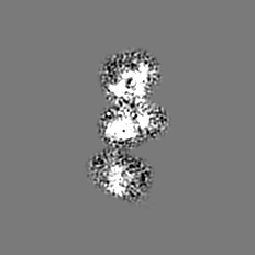
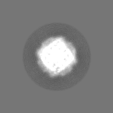
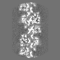
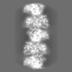
| Images |  emd_11937.png emd_11937.png | 202.2 KB | ||
| Masks | emd_11937_msk_1.map | 47.6 MB | Mask map | |
| Others | emd_11937_half_map_1.map.gzemd_11937_half_map_2.map.gz | 11.5 MB 11.5 MB | ||
| Archive directory |  http://ftp.pdbj.org/pub/emdb/structures/EMD-11937ftp://ftp.pdbj.org/pub/emdb/structures/EMD-11937 http://ftp.pdbj.org/pub/emdb/structures/EMD-11937ftp://ftp.pdbj.org/pub/emdb/structures/EMD-11937 | HTTPS FTP |
-Related structure data
| Related structure data |  7bkqMC  7bkpC  7ngaC  7nicC  7niqC C: citing same article ( M: atomic model generated by this map |
|---|---|
| Similar structure data | |
| EM raw data | EMPIAR-10653 (Title: WT MDA5-dsRNA filaments in complex with ADP / Data size: 791.8 Data #1: Unaligned multiframe micrographs of MDA5-dsRNA filaments with ADP bound [micrographs - multiframe]) |


-Links
| EMDB pages | EMDB (EBI/PDBe) / EMDataResource |
|---|---|
| Related items in Molecule of the Month |


-Map
| File | Download / File: emd_11937.map.gz / Format: CCP4 / Size: 47.6 MB / Type: IMAGE STORED AS FLOATING POINT NUMBER (4 BYTES) | ||||||||||||||||||||||||||||||||||||||||||||||||||||||||||||
|---|---|---|---|---|---|---|---|---|---|---|---|---|---|---|---|---|---|---|---|---|---|---|---|---|---|---|---|---|---|---|---|---|---|---|---|---|---|---|---|---|---|---|---|---|---|---|---|---|---|---|---|---|---|---|---|---|---|---|---|---|---|
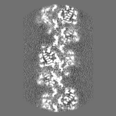
| Projections & slices | Image control
Images are generated by Spider. | ||||||||||||||||||||||||||||||||||||||||||||||||||||||||||||
| Voxel size | X=Y=Z: 1.04 Å | ||||||||||||||||||||||||||||||||||||||||||||||||||||||||||||
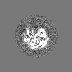
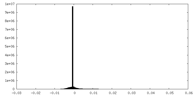
| Density |
| ||||||||||||||||||||||||||||||||||||||||||||||||||||||||||||
| Symmetry | Space group: 1 | ||||||||||||||||||||||||||||||||||||||||||||||||||||||||||||
| Details | EMDB XML:
CCP4 map header:
| ||||||||||||||||||||||||||||||||||||||||||||||||||||||||||||
 Z (Sec.)
 Y (Row.)
 X (Col.)


-Supplemental data
-Mask #1
| File | emd_11937_msk_1.map | ||||||||||||
|---|---|---|---|---|---|---|---|---|---|---|---|---|---|
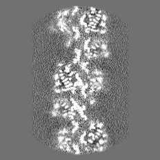
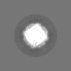
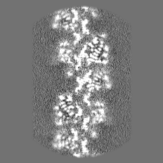
| Projections & Slices |
| ||||||||||||
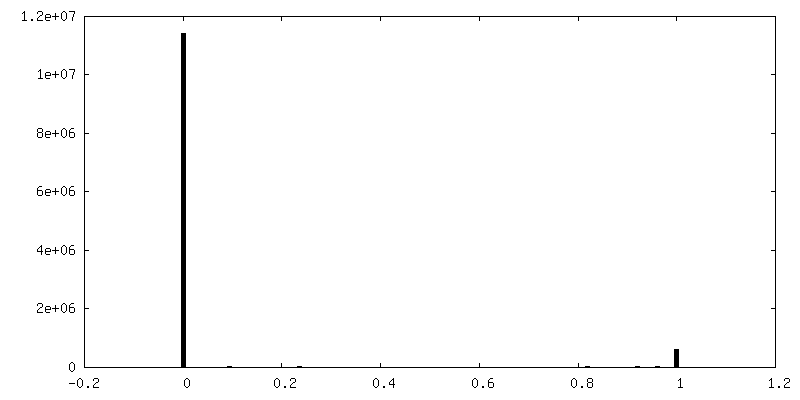
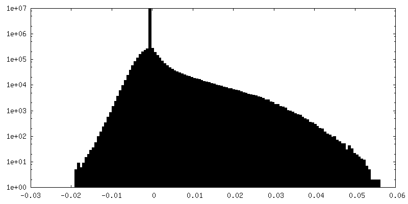
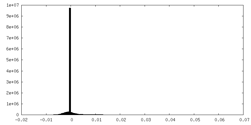
| Density Histograms |


-Half map: #2
| File | emd_11937_half_map_1.map | ||||||||||||
|---|---|---|---|---|---|---|---|---|---|---|---|---|---|
| Projections & Slices |
| ||||||||||||
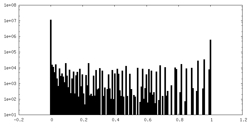
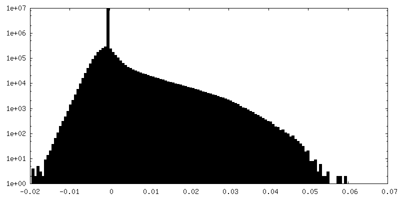
| Density Histograms |

-Half map: #1
| File | emd_11937_half_map_2.map | ||||||||||||
|---|---|---|---|---|---|---|---|---|---|---|---|---|---|
| Projections & Slices |
| ||||||||||||
| Density Histograms |

- Sample components
-Entire : MDA5-dsRNA filament in complex with ADP
| Entire | Name: MDA5-dsRNA filament in complex with ADP |
|---|---|
| Components |
|
-Supramolecule #1: MDA5-dsRNA filament in complex with ADP
| Supramolecule | Name: MDA5-dsRNA filament in complex with ADP / type: complex / ID: 1 / Parent: 0 / Macromolecule list: all |
|---|---|
| Molecular weight | Experimental: 30 kDa/nm |
-Supramolecule #2: INTERFERON-INDUCED HELICASE C DOMAIN-CONTAINING PROTEIN
| Supramolecule | Name: INTERFERON-INDUCED HELICASE C DOMAIN-CONTAINING PROTEIN type: complex / ID: 2 / Parent: 1 / Macromolecule list: #1 |
|---|---|
| Source (natural) | Organism: |
| Recombinant expression | Organism:  |
-Supramolecule #3: RNA
| Supramolecule | Name: RNA / type: complex / ID: 3 / Parent: 1 / Macromolecule list: #2-#3 |
|---|---|
| Source (natural) | Organism: Pseudomonas virus phi6 |
-Macromolecule #1: INTERFERON-INDUCED HELICASE C DOMAIN-CONTAINING PROTEIN
| Macromolecule | Name: INTERFERON-INDUCED HELICASE C DOMAIN-CONTAINING PROTEIN type: protein_or_peptide / ID: 1 / Enantiomer: LEVO / EC number: RNA helicase |
|---|---|
| Source (natural) | Organism: |
| Recombinant expression | Organism: |
| Sequence | String: LQLRPYQMEV AQPALDGKNI IICLPTGSGK TRVAVYITKD HLDKKKQASE SGKVIVLVNK VMLAEQLFRK EFNPYLKKWY RIIGLSGDTQ LKISFPEVVK SYDVIISTAQ ILENSLLNLE SGDDDGVQLS DFSLIIIDEC HHTNKEAVYN NIMRRYLKQK LRNNDLKKQN ...String: LQLRPYQMEV AQPALDGKNI IICLPTGSGK TRVAVYITKD HLDKKKQASE SGKVIVLVNK VMLAEQLFRK EFNPYLKKWY RIIGLSGDTQ LKISFPEVVK SYDVIISTAQ ILENSLLNLE SGDDDGVQLS DFSLIIIDEC HHTNKEAVYN NIMRRYLKQK LRNNDLKKQN KPAIPLPQIL GLTASPGVGA AKKQSEAEKH ILNICANLDA FTIKTVKENL GQLKHQIKEP CKKFVIADDT RENPFKEKLL EIMASIQTYC QKSPMSDFGT QHYEQWAIQM EKKAAKDGNR KDRVCAEHLR KYNEALQIND TIRMIDAYSH LETFYTDEKE KKFAVLNDSK KSLKLDETDE FLMNLFFDNK KMLKKLAENP KYENEKLIKL RNTILEQFTR SEESSRGIIF TKTRQSTYAL SQWIMENAKF AEVGVKAHHL IGAGHSSEVK PMTQTEQKEV ISKFRTGEIN LLIATTVAEE GLDIKECNIV IRYGLVTNEI AMVQARGRAR ADESTYVLVT SSGSGVTERE IVNDFREKMM YKAINRVQNM KPEEYAHKIL ELQVQSILEK KMKVKRSIAK QYNDNPSLIT LLCKNCSMLV CSGENIHVIE KMHHVNMTPE FKGLYIVREN KALQKKFADY QTNGEIICKC GQAWGTMMVH KGLDLPCLKI RNFVVNFKNN SPKKQYKKWV ELPIRFPDLD YSEYCL |
-Macromolecule #2: RNA
| Macromolecule | Name: RNA / type: rna / ID: 2 |
|---|---|
| Source (natural) | Organism: Pseudomonas virus phi6 |
| Sequence | String: UCCAUGCGCA UGACG |
-Macromolecule #3: RNA
| Macromolecule | Name: RNA / type: rna / ID: 3 |
|---|---|
| Source (natural) | Organism: Pseudomonas virus phi6 |
| Sequence | String: CGUCAUGCGC AUGGA |
-Experimental details
-Structure determination
| Method | cryo EM |
|---|---|
 Processing Processing | helical reconstruction |
| Aggregation state | filament |
-Sample preparation
| Concentration | 1 mg/mL | ||||||||||||||||||
|---|---|---|---|---|---|---|---|---|---|---|---|---|---|---|---|---|---|---|---|
| Buffer | pH: 7.7 Component:
| ||||||||||||||||||
| Grid | Model: Quantifoil R1.2/1.3 / Material: GOLD / Mesh: 300 / Support film - Material: CARBON / Support film - topology: HOLEY / Pretreatment - Type: GLOW DISCHARGE / Details: machine step 6 | ||||||||||||||||||
| Vitrification | Cryogen name: ETHANE / Chamber humidity: 100 % / Chamber temperature: 277.2 K / Instrument: FEI VITROBOT MARK IV Details: Grids were blotted for 3 s; blot force 10, 5, 0, -5, -10. |
- Electron microscopy
| Microscope | FEI TITAN KRIOS |
|---|---|
| Specialist optics | Phase plate: OTHER / Spherical aberration corrector: None / Energy filter - Slit width: 20 eV |
| Image recording | Film or detector model: GATAN K2 SUMMIT (4k x 4k) / Detector mode: COUNTING / Digitization - Dimensions - Width: 3838 pixel / Digitization - Dimensions - Height: 3710 pixel / Digitization - Frames/image: 1-40 / Number grids imaged: 1 / Number real images: 4680 / Average electron dose: 1.1125 e/Å2 |
| Electron beam | Acceleration voltage: 300 kV / Electron source:  FIELD EMISSION GUN FIELD EMISSION GUN |
| Electron optics | Illumination mode: FLOOD BEAM / Imaging mode: BRIGHT FIELD / Nominal defocus max: 2.7 µm / Nominal defocus min: 1.8 µm |
| Sample stage | Specimen holder model: FEI TITAN KRIOS AUTOGRID HOLDER / Cooling holder cryogen: NITROGEN |
| Experimental equipment |  Model: Titan Krios / Image courtesy: FEI Company |