 ムービー
ムービー コントローラー

[日本語] English
 万見
万見- EMDB-1108: Structural basis of pore formation by the bacterial toxin pneumolysin. -
+ データを開く

- 基本情報
基本情報
| 登録情報 | データベース: EMDB / ID: EMD-1108 | |||||||||
|---|---|---|---|---|---|---|---|---|---|---|
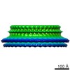
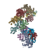
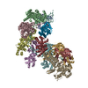
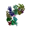
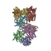
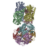
| タイトル | Structural basis of pore formation by the bacterial toxin pneumolysin. | |||||||||
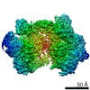
 マップデータ マップデータ | This 3D map is the weighted average of two pneumolysin pore maps of different sizes. | |||||||||
 試料 試料 |
| |||||||||
| 機能・相同性 |  機能・相同性情報 機能・相同性情報hemolysis in another organism / cholesterol binding / toxin activity / membrane => GO:0016020 / host cell plasma membrane / extracellular region / membrane 類似検索 - 分子機能 | |||||||||
| 生物種 | synthetic construct (人工物) /   Streptococcus pneumoniae (肺炎レンサ球菌) Streptococcus pneumoniae (肺炎レンサ球菌) | |||||||||
| 手法 | 単粒子再構成法 / クライオ電子顕微鏡法 / 解像度: 28.0 Å | |||||||||
 データ登録者 データ登録者 | Tilley SJ / Orlova EV / Gilbert RJ / Andrew PW / Saibil HR | |||||||||
 引用 引用 | ジャーナル: Cell / 年: 2005 タイトル: Structural basis of pore formation by the bacterial toxin pneumolysin. 著者: Sarah J Tilley / Elena V Orlova / Robert J C Gilbert / Peter W Andrew / Helen R Saibil /  要旨: The bacterial toxin pneumolysin is released as a soluble monomer that kills target cells by assembling into large oligomeric rings and forming pores in cholesterol-containing membranes. Using cryo-EM ...The bacterial toxin pneumolysin is released as a soluble monomer that kills target cells by assembling into large oligomeric rings and forming pores in cholesterol-containing membranes. Using cryo-EM and image processing, we have determined the structures of membrane-surface bound (prepore) and inserted-pore oligomer forms, providing a direct observation of the conformational transition into the pore form of a cholesterol-dependent cytolysin. In the pore structure, the domains of the monomer separate and double over into an arch, forming a wall sealing the bilayer around the pore. This transformation is accomplished by substantial refolding of two of the four protein domains along with deformation of the membrane. Extension of protein density into the bilayer supports earlier predictions that the protein inserts beta hairpins into the membrane. With an oligomer size of up to 44 subunits in the pore, this assembly creates a transmembrane channel 260 A in diameter lined by 176 beta strands. | |||||||||
| 履歴 |
|
- 構造の表示
構造の表示
| ムービー |
ムービービューア |
|---|---|
| 構造ビューア | EMマップ: SurfViewMolmilJmol/JSmol |
| 添付画像 |
- ダウンロードとリンク
-EMDBアーカイブ
| マップデータ | emd_1108.map.gz | 295.9 KB | EMDBマップデータ形式 | |
|---|---|---|---|---|
| ヘッダ (付随情報) | emd-1108-v30.xmlemd-1108.xml | 12.3 KB 12.3 KB | 表示 表示 | EMDBヘッダ |
| 画像 |  1108.gif 1108.gif | 133.8 KB | ||
| アーカイブディレクトリ |  http://ftp.pdbj.org/pub/emdb/structures/EMD-1108ftp://ftp.pdbj.org/pub/emdb/structures/EMD-1108 http://ftp.pdbj.org/pub/emdb/structures/EMD-1108ftp://ftp.pdbj.org/pub/emdb/structures/EMD-1108 | HTTPS FTP |
-検証レポート
| 文書・要旨 | emd_1108_validation.pdf.gz | 189.8 KB | 表示 | EMDB検証レポート |
|---|---|---|---|---|
| 文書・詳細版 | emd_1108_full_validation.pdf.gz | 188.9 KB | 表示 | |
| XML形式データ | emd_1108_validation.xml.gz | 5.7 KB | 表示 | |
| アーカイブディレクトリ | https://ftp.pdbj.org/pub/emdb/validation_reports/EMD-1108ftp://ftp.pdbj.org/pub/emdb/validation_reports/EMD-1108 | HTTPS FTP |
-関連構造データ


-リンク
| EMDBのページ | EMDB (EBI/PDBe) / EMDataResource |
|---|
-マップ
| ファイル | ダウンロード / ファイル: emd_1108.map.gz / 形式: CCP4 / 大きさ: 15.3 MB / タイプ: IMAGE STORED AS FLOATING POINT NUMBER (4 BYTES) | ||||||||||||||||||||||||||||||||||||||||||||||||||||||||||||||||||||
|---|---|---|---|---|---|---|---|---|---|---|---|---|---|---|---|---|---|---|---|---|---|---|---|---|---|---|---|---|---|---|---|---|---|---|---|---|---|---|---|---|---|---|---|---|---|---|---|---|---|---|---|---|---|---|---|---|---|---|---|---|---|---|---|---|---|---|---|---|---|
| 注釈 | This 3D map is the weighted average of two pneumolysin pore maps of different sizes. | ||||||||||||||||||||||||||||||||||||||||||||||||||||||||||||||||||||
| ボクセルのサイズ | X=Y=Z: 3.5 Å | ||||||||||||||||||||||||||||||||||||||||||||||||||||||||||||||||||||
| 密度 |
| ||||||||||||||||||||||||||||||||||||||||||||||||||||||||||||||||||||
| 対称性 | 空間群: 1 | ||||||||||||||||||||||||||||||||||||||||||||||||||||||||||||||||||||
| 詳細 | EMDB XML:
CCP4マップ ヘッダ情報:
| ||||||||||||||||||||||||||||||||||||||||||||||||||||||||||||||||||||
-添付データ
- 試料の構成要素
-全体 : Pneumolysin
| 全体 | 名称: Pneumolysin |
|---|---|
| 要素 |
|
-超分子 #1000: Pneumolysin
| 超分子 | 名称: Pneumolysin / タイプ: sample / ID: 1000 / Number unique components: 2 |
|---|
-超分子 #1: Phosphatidylcholine-cholesterol lipid bilayer
| 超分子 | 名称: Phosphatidylcholine-cholesterol lipid bilayer / タイプ: organelle_or_cellular_component / ID: 1 / 組換発現: No / データベース: NCBI |
|---|---|
| 由来(天然) | 生物種: synthetic construct (人工物) / 別称: membrane |
-分子 #1: Pneumolysin
| 分子 | 名称: Pneumolysin / タイプ: protein_or_peptide / ID: 1 / 組換発現: Yes |
|---|---|
| 由来(天然) | 生物種: Streptococcus pneumoniae (肺炎レンサ球菌) / 細胞中の位置: cytoplasm |
| 組換発現 | 生物種: |
| 配列 | InterPro: Thiol-activated cytolysin |
-実験情報
-構造解析
| 手法 | クライオ電子顕微鏡法 |
|---|---|
 解析 解析 | 単粒子再構成法 |
| 試料の集合状態 | particle |
-試料調製
| 濃度 | 0.05 mg/mL |
|---|---|
| 緩衝液 | pH: 6.95 / 詳細: 8 mM Na2HPO4, 1.5 mM KH2PO4, 2.5 mM KCl, 0.25M NaCl |
| グリッド | 詳細: holey carbon 400 mesh copper grid, glow discharged using positive charge |
| 凍結 | 凍結剤: ETHANE / チャンバー内湿度: 96 % / チャンバー内温度: 100 K / 装置: HOMEMADE PLUNGER / 詳細: Vitrification instrument: home made plunger 手法: Grids were blotted for approximately 3 seconds and allowed to drain vertically for 5 seconds before plunging. |
- 電子顕微鏡法
| 顕微鏡 | FEI TECNAI F20 |
|---|---|
| 温度 | 最低: 100 K / 最高: 100 K / 平均: 100 K |
| アライメント法 | Legacy - 非点収差: corrected at 150,000 magnification |
| 撮影 | カテゴリ: FILM / フィルム・検出器のモデル: KODAK SO-163 FILM / デジタル化 - スキャナー: ZEISS SCAI / デジタル化 - サンプリング間隔: 7 µm / 実像数: 135 / 平均電子線量: 20 e/Å2 / 詳細: After scanning images were averaged 2x2. / Od range: 1 / ビット/ピクセル: 8 |
| 電子線 | 加速電圧: 200 kV / 電子線源:  FIELD EMISSION GUN FIELD EMISSION GUN |
| 電子光学系 | 照射モード: FLOOD BEAM / 撮影モード: BRIGHT FIELD / Cs: 2.0 mm / 最大 デフォーカス(公称値): 3.2 µm / 最小 デフォーカス(公称値): 1.1 µm / 倍率(公称値): 40000 |
| 試料ステージ | 試料ホルダー: Side entry / 試料ホルダーモデル: GATAN LIQUID NITROGEN |
| 実験機器 |  モデル: Tecnai F20 / 画像提供: FEI Company |
+画像解析
-原子モデル構築 1
| 初期モデル | PDB ID: |
|---|---|
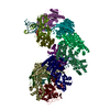
| 詳細 | The 1pfo structure was separated into six rigid bodies: domain 1 (91-172, 231-272, 354-373), domain 2 upper (53-62, 83-90, 374-382), domain 2 lower (63-82, 382-390), domain 3 (177-186, 221-230, 273-283, 316-355), domain 3 hairpins (187-220, 284-315), and domain 4 (391-500). These rigid bodies were fitted manually using the software O and pymol. |
| 精密化 | プロトコル: RIGID BODY FIT |
| 得られたモデル | 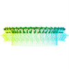 PDB-2bk1: |

