- EMDB-11058: Human ER Membrane protein Complex (EMC) -
+
Open data
ID or keywords:
Loading...
-
Basic information
Entry
Database: EMDB / ID: EMD-11058
Title
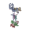
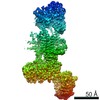
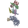
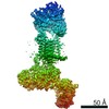
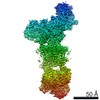
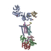
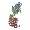
Human ER Membrane protein Complex (EMC)
Map data
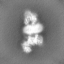
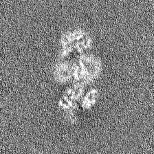
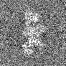
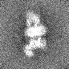
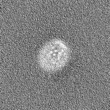
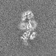
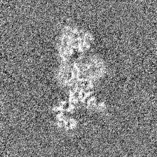
3D reconstruction of the human ER-Membrane Complex produced by single particle cryo-electron microscopy.
Sample
Complex: Human ER Membrane protein Complex (EMC)
Function / homology
Function and homology information
extrinsic component of endoplasmic reticulum membrane / EMC complex / : / omegasome membrane / protein insertion into ER membrane by stop-transfer membrane-anchor sequence / magnesium ion transport / tail-anchored membrane protein insertion into ER membrane / Miscellaneous transport and binding events / cobalt ion transmembrane transporter activity / ferrous iron transmembrane transporter activity ...extrinsic component of endoplasmic reticulum membrane / EMC complex / : / omegasome membrane / protein insertion into ER membrane by stop-transfer membrane-anchor sequence / magnesium ion transport / tail-anchored membrane protein insertion into ER membrane / Miscellaneous transport and binding events / cobalt ion transmembrane transporter activity / ferrous iron transmembrane transporter activity / copper ion transport / magnesium ion transmembrane transporter activity / RHOA GTPase cycle / autophagosome assembly / positive regulation of endothelial cell proliferation / positive regulation of endothelial cell migration / positive regulation of angiogenesis / carbohydrate binding / angiogenesis / early endosome membrane / early endosome / Golgi membrane / apoptotic process / endoplasmic reticulum membrane / Golgi apparatus / endoplasmic reticulum / protein-containing complex / extracellular region / membrane / plasma membrane / cytoplasm Similarity search - Function
: / EMC1 N-terminal beta-propeller domain / ER membrane protein complex subunit 8/9 / : / Uncharacterised protein family (UPF0172) / EMC2 TPR-like repeat domain / TMEM85/ER membrane protein complex subunit 4 / ER membrane protein complex subunit 4 / ER membrane protein complex subunit 7, beta-sandwich domain / ER membrane protein complex subunit 7 ...: / EMC1 N-terminal beta-propeller domain / ER membrane protein complex subunit 8/9 / : / Uncharacterised protein family (UPF0172) / EMC2 TPR-like repeat domain / TMEM85/ER membrane protein complex subunit 4 / ER membrane protein complex subunit 4 / ER membrane protein complex subunit 7, beta-sandwich domain / ER membrane protein complex subunit 7 / ER membrane protein complex subunit 7, beta-sandwich domain / ER membrane protein complex subunit 6 / ER membrane protein complex subunit 3 / ER membrane protein complex subunit 1, C-terminal / Membrane magnesium transporter / ER membrane protein complex subunit 1 / ER membrane protein complex subunit 6-like / EMC6 / ER membrane protein complex subunit 1, second beta-propeller / Membrane magnesium transporter / ER membrane protein complex subunit 10 / ER membrane protein complex subunit 2-like / Integral membrane protein EMC3/TMCO1-like / Integral membrane protein EMC3/TMCO1-like / Integral membrane protein DUF106 / Carbohydrate-binding-like fold / Quinoprotein alcohol dehydrogenase-like superfamily / TPR repeat region circular profile. / TPR repeat profile. / MPN domain / MPN domain profile. / Tetratricopeptide repeats / Tetratricopeptide repeat / Tetratricopeptide-like helical domain superfamily / WD40/YVTN repeat-like-containing domain superfamily Similarity search - Domain/homology
ER membrane protein complex subunit 2 / ER membrane protein complex subunit 4 / ER membrane protein complex subunit 10 / ER membrane protein complex subunit 5 / ER membrane protein complex subunit 1 / ER membrane protein complex subunit 6 / Endoplasmic reticulum membrane protein complex subunit 7 / ER membrane protein complex subunit 3 / ER membrane protein complex subunit 9 Similarity search - Component
Biological species
Homo sapiens (human)
Method
single particle reconstruction / cryo EM / Resolution: 6.4 Å
Journal: Elife / Year: 2020 Title: The architecture of EMC reveals a path for membrane protein insertion. Authors: John P O'Donnell / Ben P Phillips / Yuichi Yagita / Szymon Juszkiewicz / Armin Wagner / Duccio Malinverni / Robert J Keenan / Elizabeth A Miller / Ramanujan S Hegde / Abstract: Approximately 25% of eukaryotic genes code for integral membrane proteins that are assembled at the endoplasmic reticulum. An abundant and widely conserved multi-protein complex termed EMC has been ...Approximately 25% of eukaryotic genes code for integral membrane proteins that are assembled at the endoplasmic reticulum. An abundant and widely conserved multi-protein complex termed EMC has been implicated in membrane protein biogenesis, but its mechanism of action is poorly understood. Here, we define the composition and architecture of human EMC using biochemical assays, crystallography of individual subunits, site-specific photocrosslinking, and cryo-EM reconstruction. Our results suggest that EMC's cytosolic domain contains a large, moderately hydrophobic vestibule that can bind a substrate's transmembrane domain (TMD). The cytosolic vestibule leads into a lumenally-sealed, lipid-exposed intramembrane groove large enough to accommodate a single substrate TMD. A gap between the cytosolic vestibule and intramembrane groove provides a potential path for substrate egress from EMC. These findings suggest how EMC facilitates energy-independent membrane insertion of TMDs, explain why only short lumenal domains are translocated by EMC, and constrain models of EMC's proposed chaperone function.
History
Deposition
May 20, 2020
-
Header (metadata) release
Jun 17, 2020
-
Map release
Jun 17, 2020
-
Update
Jun 17, 2020
-
Current status
Jun 17, 2020
Processing site: PDBe / Status: Released
-
Structure visualization
Movie
Surface view with section colored by density value
This sample was purified from suspension adapted human HEK293 cells and was monodisperse on EM grids.
-
Electron microscopy
Microscope
FEI TITAN KRIOS
Temperature
Min: 84.0 K / Max: 93.0 K
Specialist optics
Phase plate: VOLTA PHASE PLATE / Energy filter - Slit width: 20 eV
Image recording
Film or detector model: GATAN K2 SUMMIT (4k x 4k) / Detector mode: COUNTING / Digitization - Dimensions - Width: 3710 pixel / Digitization - Dimensions - Height: 3710 pixel / Digitization - Sampling interval: 5.0 µm / Digitization - Frames/image: 1-44 / Number grids imaged: 5 / Number real images: 11626 / Average exposure time: 11.0 sec. / Average electron dose: 39.36 e/Å2 Details: Data collected across 5 independent sessions using two different FEI Titan Krios Microscopes, both equipped with a Gatan K2 Summit detector and volta phase plate. Volta phase plate used ...Details: Data collected across 5 independent sessions using two different FEI Titan Krios Microscopes, both equipped with a Gatan K2 Summit detector and volta phase plate. Volta phase plate used without charging between exposures. Phase plate evolution varied between different microscopes used to collect data but positions were shifted once phase evolution progressed past 120 degrees.
Electron beam
Acceleration voltage: 300 kV / Electron source: FIELD EMISSION GUN
Type of model: INSILICO MODEL / In silico model: Ab initio model generation in CryoSPARC. Details: 4 classes generated and best class used as a initial model for refinement.
Final reconstruction
Number classes used: 1 / Applied symmetry - Point group: C1 (asymmetric) / Algorithm: FOURIER SPACE / Resolution.type: BY AUTHOR / Resolution: 6.4 Å / Resolution method: FSC 0.143 CUT-OFF / Software - Name: cryoSPARC (ver. 2.12.4) Software - details: Local resolution estimation and filtering Details: Resolution calculated from half maps produced during local resolution estimation in CryoSPARC following non-uniform refinement. Number images used: 167294
Initial angle assignment
Type: MAXIMUM LIKELIHOOD / Software - Name: cryoSPARC (ver. 2.12.4) / Software - details: Ab Initio Model generation
Number classes: 4 / Avg.num./class: 85780 / Software - Name: cryoSPARC (ver. 2.12.4) / Software - details: Ab Initio 3D classification / Details: Ab Initio classification in CryoSPARC
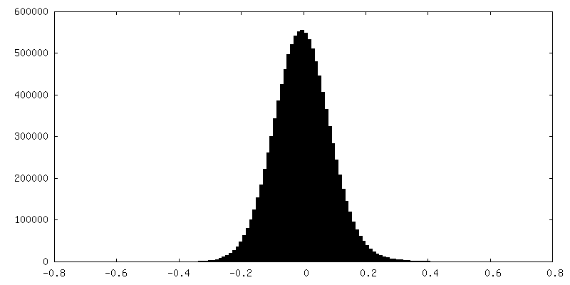
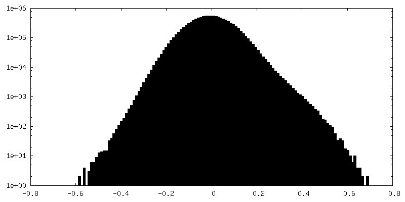
FSC plot (resolution estimation)
+
About Yorodumi
-
News
-
Feb 9, 2022. New format data for meta-information of EMDB entries
New format data for meta-information of EMDB entries
Version 3 of the EMDB header file is now the official format.
The previous official version 1.9 will be removed from the archive.
In the structure databanks used in Yorodumi, some data are registered as the other names, "COVID-19 virus" and "2019-nCoV". Here are the details of the virus and the list of structure data.
Jan 31, 2019. EMDB accession codes are about to change! (news from PDBe EMDB page)
EMDB accession codes are about to change! (news from PDBe EMDB page)
The allocation of 4 digits for EMDB accession codes will soon come to an end. Whilst these codes will remain in use, new EMDB accession codes will include an additional digit and will expand incrementally as the available range of codes is exhausted. The current 4-digit format prefixed with “EMD-” (i.e. EMD-XXXX) will advance to a 5-digit format (i.e. EMD-XXXXX), and so on. It is currently estimated that the 4-digit codes will be depleted around Spring 2019, at which point the 5-digit format will come into force.
The EM Navigator/Yorodumi systems omit the EMD- prefix.
Related info.:Q: What is EMD? / ID/Accession-code notation in Yorodumi/EM Navigator
Yorodumi is a browser for structure data from EMDB, PDB, SASBDB, etc.
This page is also the successor to EM Navigator detail page, and also detail information page/front-end page for Omokage search.
The word "yorodu" (or yorozu) is an old Japanese word meaning "ten thousand". "mi" (miru) is to see.
Related info.:EMDB / PDB / SASBDB / Comparison of 3 databanks / Yorodumi Search / Aug 31, 2016. New EM Navigator & Yorodumi / Yorodumi Papers / Jmol/JSmol / Function and homology information / Changes in new EM Navigator and Yorodumi
 Movie
Movie Controller


 Open data
 Basic information
 Map data
 Sample
 Function and homology information
 Homo sapiens (human)
 Authors
 United Kingdom, 3 items
  Citation
 Structure visualization
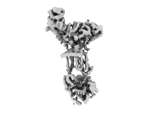
 Downloads & links
 emd_11058.png
 http://ftp.pdbj.org/pub/emdb/structures/EMD-11058


 Z (Sec.)
 Y (Row.)
 X (Col.)


 Sample components
 Processing
 Electron microscopy
 FIELD EMISSION GUN

