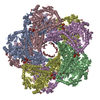
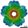
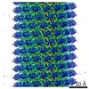
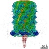
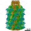
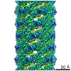
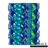
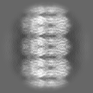
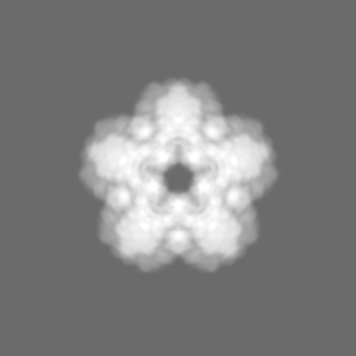
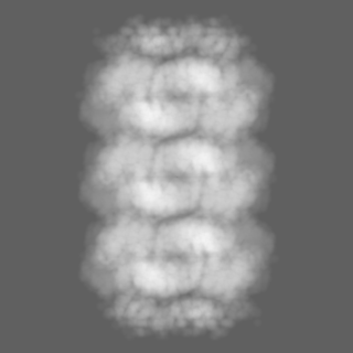
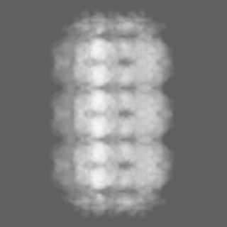
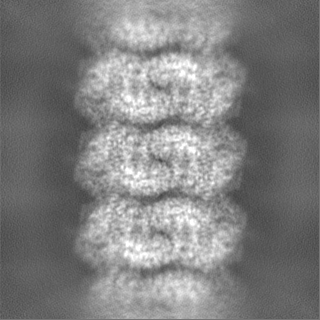
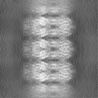
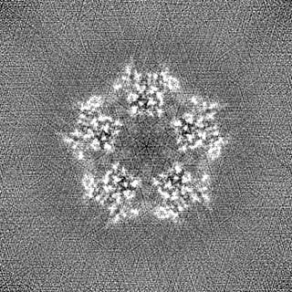
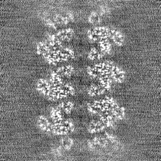
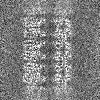
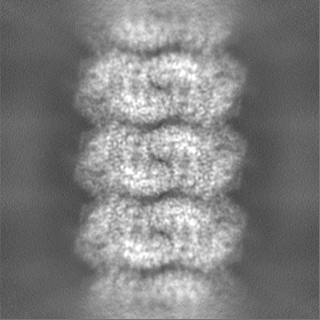
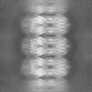
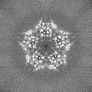
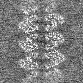
Journal: Proc Natl Acad Sci U S A / Year: 2021 Title: Supramolecular assembly of the LdcI upon acid stress. Authors: Matthew Jessop / Clarissa Liesche / Jan Felix / Ambroise Desfosses / Megghane Baulard / Virgile Adam / Angélique Fraudeau / Karine Huard / Grégory Effantin / Jean-Philippe Kleman / Maria ...Authors: Matthew Jessop / Clarissa Liesche / Jan Felix / Ambroise Desfosses / Megghane Baulard / Virgile Adam / Angélique Fraudeau / Karine Huard / Grégory Effantin / Jean-Philippe Kleman / Maria Bacia-Verloop / Dominique Bourgeois / Irina Gutsche / Abstract: Pathogenic and commensal bacteria often have to resist the harsh acidity of the host stomach. The inducible lysine decarboxylase LdcI buffers the cytosol and the local extracellular environment to ...Pathogenic and commensal bacteria often have to resist the harsh acidity of the host stomach. The inducible lysine decarboxylase LdcI buffers the cytosol and the local extracellular environment to ensure enterobacterial survival at low pH. Here, we investigate the acid stress-response regulation of LdcI by combining biochemical and biophysical characterization with negative stain and cryoelectron microscopy (cryo-EM) and wide-field and superresolution fluorescence imaging. Due to deleterious effects of fluorescent protein fusions on native LdcI decamers, we opt for three-dimensional localization of nanobody-labeled endogenous wild-type LdcI in acid-stressed cells and show that it organizes into distinct patches at the cell periphery. Consistent with recent hypotheses that in vivo clustering of metabolic enzymes often reflects their polymerization as a means of stimulus-induced regulation, we show that LdcI assembles into filaments in vitro at physiologically relevant low pH. We solve the structures of these filaments and of the LdcI decamer formed at neutral pH by cryo-EM and reveal the molecular determinants of LdcI polymerization, confirmed by mutational analysis. Finally, we propose a model for LdcI function inside the enterobacterial cell, providing a structural and mechanistic basis for further investigation of the role of its supramolecular organization in the acid stress response.
History
Deposition
Apr 10, 2020
-
Header (metadata) release
Jan 13, 2021
-
Map release
Jan 13, 2021
-
Update
Apr 9, 2025
-
Current status
Apr 9, 2025
Processing site: PDBe / Status: Released
-
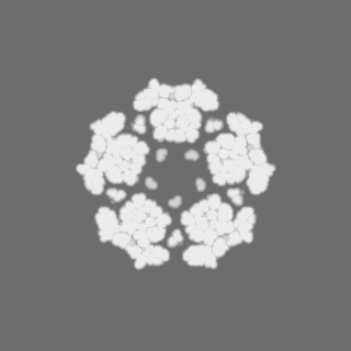
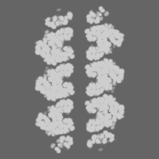
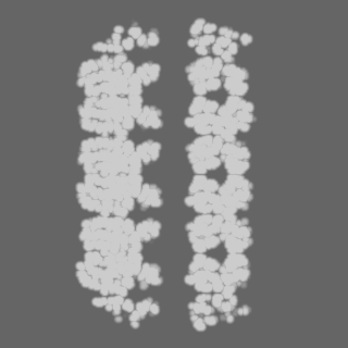
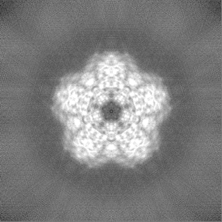
Structure visualization
Movie
Surface view with section colored by density value
Model: Quantifoil R2/1 / Material: COPPER / Mesh: 300 / Support film - Material: CARBON / Support film - topology: HOLEY / Pretreatment - Type: GLOW DISCHARGE / Pretreatment - Time: 40 sec.
Vitrification
Cryogen name: ETHANE / Chamber humidity: 100 % / Instrument: FEI VITROBOT MARK IV
-
Electron microscopy
Microscope
TFS KRIOS
Specialist optics
Energy filter - Name: GIF Bioquantum / Energy filter - Slit width: 20 eV
Image recording
Film or detector model: GATAN K2 SUMMIT (4k x 4k) / Detector mode: COUNTING / Number real images: 2564 / Average exposure time: 6.0 sec. / Average electron dose: 29.3 e/Å2
Electron beam
Acceleration voltage: 300 kV / Electron source: FIELD EMISSION GUN
Details: Initial model generated by manually stacking three LdcI decamers (PDB ID: 3N75) in Chimera and low-pass filtering the resulting LdcI-stack to 40 A.
Final reconstruction
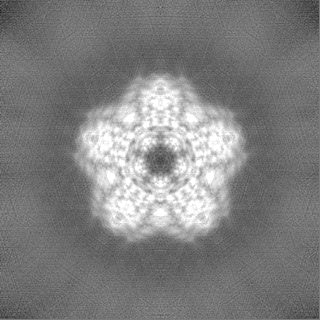
Applied symmetry - Point group: D5 (2x5 fold dihedral) / Resolution.type: BY AUTHOR / Resolution: 3.28 Å / Resolution method: FSC 0.143 CUT-OFF / Software - Name: cryoSPARC / Number images used: 15157
Initial angle assignment
Type: MAXIMUM LIKELIHOOD / Software - Name: cryoSPARC
Final angle assignment
Type: MAXIMUM LIKELIHOOD / Software - Name: cryoSPARC
In the structure databanks used in Yorodumi, some data are registered as the other names, "COVID-19 virus" and "2019-nCoV". Here are the details of the virus and the list of structure data.
Jan 31, 2019. EMDB accession codes are about to change! (news from PDBe EMDB page)
EMDB accession codes are about to change! (news from PDBe EMDB page)
The allocation of 4 digits for EMDB accession codes will soon come to an end. Whilst these codes will remain in use, new EMDB accession codes will include an additional digit and will expand incrementally as the available range of codes is exhausted. The current 4-digit format prefixed with “EMD-” (i.e. EMD-XXXX) will advance to a 5-digit format (i.e. EMD-XXXXX), and so on. It is currently estimated that the 4-digit codes will be depleted around Spring 2019, at which point the 5-digit format will come into force.
The EM Navigator/Yorodumi systems omit the EMD- prefix.
Related info.:Q: What is EMD? / ID/Accession-code notation in Yorodumi/EM Navigator
Yorodumi is a browser for structure data from EMDB, PDB, SASBDB, etc.
This page is also the successor to EM Navigator detail page, and also detail information page/front-end page for Omokage search.
The word "yorodu" (or yorozu) is an old Japanese word meaning "ten thousand". "mi" (miru) is to see.
Related info.:EMDB / PDB / SASBDB / Comparison of 3 databanks / Yorodumi Search / Aug 31, 2016. New EM Navigator & Yorodumi / Yorodumi Papers / Jmol/JSmol / Function and homology information / Changes in new EM Navigator and Yorodumi
 Movie
Movie Controller


 Open data
 Basic information
 Map data
 Sample
 Keywords
 Function and homology information
 Authors
 Citation
 Structure visualization
 Downloads & links
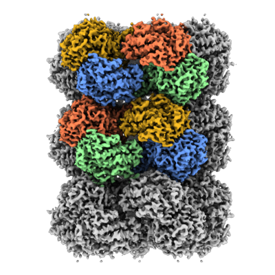 emd_10850.png
 http://ftp.pdbj.org/pub/emdb/structures/EMD-10850

 Z (Sec.)
 Y (Row.)
 X (Col.)


 Sample components
 Processing
 Electron microscopy
 FIELD EMISSION GUN