Movie
Movie Controller


+ Open data

- Basic information
| Entry | Database: EMDB / ID: EMD-0643 | |||||||||||||||
|---|---|---|---|---|---|---|---|---|---|---|---|---|---|---|---|---|
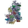
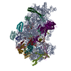
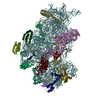
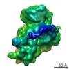
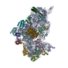
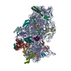
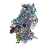
| Title | 30S initiation complex | |||||||||||||||
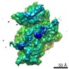
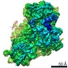
 Map data Map data | 30S IC | |||||||||||||||
 Sample Sample |
| |||||||||||||||
 Keywords Keywords | 30S Initiation complex with IF1 / IF2 and P/I tRNA / RIBOSOME | |||||||||||||||
| Function / homology |  Function and homology information Function and homology informationtranslation initiation factor activity / small ribosomal subunit / small ribosomal subunit rRNA binding / cytosolic small ribosomal subunit / cytoplasmic translation / tRNA binding / rRNA binding / structural constituent of ribosome / ribosome / translation ...translation initiation factor activity / small ribosomal subunit / small ribosomal subunit rRNA binding / cytosolic small ribosomal subunit / cytoplasmic translation / tRNA binding / rRNA binding / structural constituent of ribosome / ribosome / translation / ribonucleoprotein complex / mRNA binding / GTPase activity / GTP binding / cytoplasm / cytosol Similarity search - Function | |||||||||||||||
| Biological species |  | |||||||||||||||
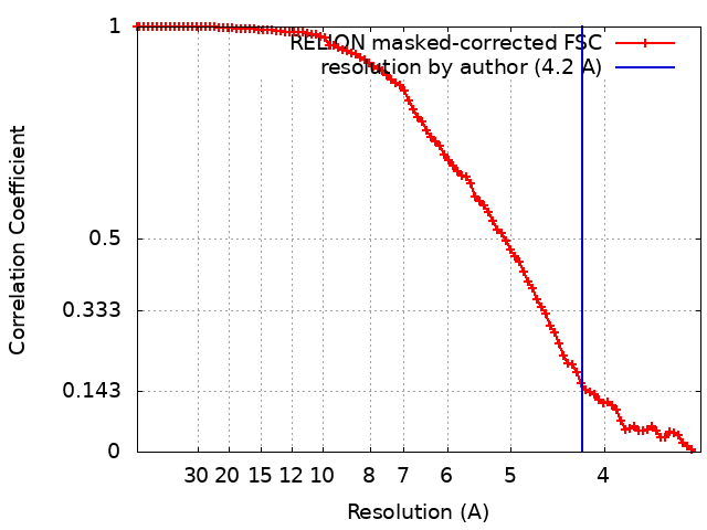
| Method | single particle reconstruction / cryo EM / Resolution: 4.2 Å | |||||||||||||||
 Authors Authors | Frank J / Gonzalez Jr RL | |||||||||||||||
| Funding support |  United States, 4 items United States, 4 items
| |||||||||||||||
 Citation Citation | Journal: Nature / Year: 2019 Title: Late steps in bacterial translation initiation visualized using time-resolved cryo-EM. Authors: Sandip Kaledhonkar / Ziao Fu / Kelvin Caban / Wen Li / Bo Chen / Ming Sun / Ruben L Gonzalez / Joachim Frank / Abstract: The initiation of bacterial translation involves the tightly regulated joining of the 50S ribosomal subunit to an initiator transfer RNA (fMet-tRNA)-containing 30S ribosomal initiation complex to ...The initiation of bacterial translation involves the tightly regulated joining of the 50S ribosomal subunit to an initiator transfer RNA (fMet-tRNA)-containing 30S ribosomal initiation complex to form a 70S initiation complex, which subsequently matures into a 70S elongation-competent complex. Rapid and accurate formation of the 70S initiation complex is promoted by initiation factors, which must dissociate from the 30S initiation complex before the resulting 70S elongation-competent complex can begin the elongation of translation. Although comparisons of the structures of the 30S and 70S initiation complexes have revealed that the ribosome, initiation factors and fMet-tRNA can acquire different conformations in these complexes, the timing of conformational changes during formation of the 70S initiation complex, the structures of any intermediates formed during these rearrangements, and the contributions that these dynamics might make to the mechanism and regulation of initiation remain unknown. Moreover, the absence of a structure of the 70S elongation-competent complex formed via an initiation-factor-catalysed reaction has precluded an understanding of the rearrangements to the ribosome, initiation factors and fMet-tRNA that occur during maturation of a 70S initiation complex into a 70S elongation-competent complex. Here, using time-resolved cryogenic electron microscopy, we report the near-atomic-resolution view of how a time-ordered series of conformational changes drive and regulate subunit joining, initiation factor dissociation and fMet-tRNA positioning during formation of the 70S elongation-competent complex. Our results demonstrate the power of time-resolved cryogenic electron microscopy to determine how a time-ordered series of conformational changes contribute to the mechanism and regulation of one of the most fundamental processes in biology. | |||||||||||||||
| History |
|
- Structure visualization
| Movie |
Movie viewer |
|---|---|
| Structure viewer | EM map: SurfViewMolmilJmol/JSmol |
| Supplemental images |

- Downloads & links
-EMDB archive
| Map data | emd_0643.map.gz | 5.4 MB | EMDB map data format | |
|---|---|---|---|---|
| Header (meta data) | emd-0643-v30.xmlemd-0643.xml | 34.3 KB 34.3 KB | Display Display | EMDB header |
| FSC (resolution estimation) | emd_0643_fsc.xml | 9.1 KB | Display | FSC data file |
| Images | 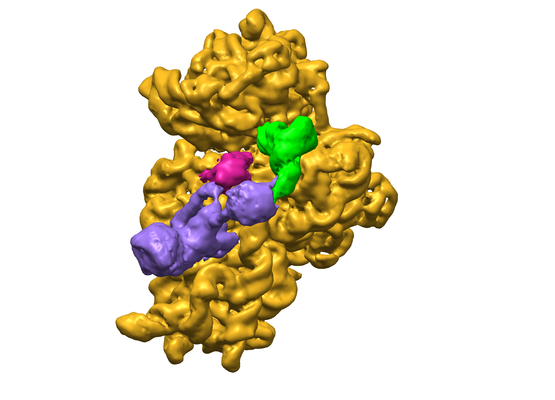 emd_0643.png emd_0643.png | 123.1 KB | ||
| Filedesc metadata | emd-0643.cif.gz | 9 KB | ||
| Archive directory |  http://ftp.pdbj.org/pub/emdb/structures/EMD-0643ftp://ftp.pdbj.org/pub/emdb/structures/EMD-0643 http://ftp.pdbj.org/pub/emdb/structures/EMD-0643ftp://ftp.pdbj.org/pub/emdb/structures/EMD-0643 | HTTPS FTP |
-Related structure data
| Related structure data |  6o7kMC  0661C  0662C 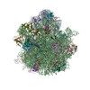 6o9jC  6o9kC M: atomic model generated by this map C: citing same article ( |
|---|---|
| Similar structure data |

-Links
| EMDB pages | EMDB (EBI/PDBe) / EMDataResource |
|---|---|
| Related items in Molecule of the Month |


-Map
| File | Download / File: emd_0643.map.gz / Format: CCP4 / Size: 64 MB / Type: IMAGE STORED AS FLOATING POINT NUMBER (4 BYTES) | ||||||||||||||||||||||||||||||||||||||||||||||||||||||||||||
|---|---|---|---|---|---|---|---|---|---|---|---|---|---|---|---|---|---|---|---|---|---|---|---|---|---|---|---|---|---|---|---|---|---|---|---|---|---|---|---|---|---|---|---|---|---|---|---|---|---|---|---|---|---|---|---|---|---|---|---|---|---|
| Annotation | 30S IC | ||||||||||||||||||||||||||||||||||||||||||||||||||||||||||||
| Projections & slices | Image control
Images are generated by Spider. | ||||||||||||||||||||||||||||||||||||||||||||||||||||||||||||
| Voxel size | X=Y=Z: 1.66 Å | ||||||||||||||||||||||||||||||||||||||||||||||||||||||||||||
| Density |
| ||||||||||||||||||||||||||||||||||||||||||||||||||||||||||||
| Symmetry | Space group: 1 | ||||||||||||||||||||||||||||||||||||||||||||||||||||||||||||
| Details | EMDB XML:
CCP4 map header:
| ||||||||||||||||||||||||||||||||||||||||||||||||||||||||||||
 Z (Sec.)
 Y (Row.)
 X (Col.)


-Supplemental data
- Sample components
+Entire : 70S elongation competent ribosome
+Supramolecule #1: 70S elongation competent ribosome
+Macromolecule #1: Translation initiation factor IF-1
+Macromolecule #2: Translation initiation factor IF-2
+Macromolecule #4: 30S ribosomal protein S17
+Macromolecule #5: 30S ribosomal protein S10
+Macromolecule #6: 30S ribosomal protein S11
+Macromolecule #7: 30S ribosomal protein S12
+Macromolecule #8: 30S ribosomal protein S13
+Macromolecule #9: 30S ribosomal protein S14
+Macromolecule #10: 30S ribosomal protein S15
+Macromolecule #11: 30S ribosomal protein S16
+Macromolecule #12: 30S ribosomal protein S18
+Macromolecule #13: 30S ribosomal protein S19
+Macromolecule #14: 30S ribosomal protein S2
+Macromolecule #15: 30S ribosomal protein S20
+Macromolecule #16: 30S ribosomal protein S21
+Macromolecule #17: 30S ribosomal protein S3
+Macromolecule #18: 30S ribosomal protein S4
+Macromolecule #19: 30S ribosomal protein S5
+Macromolecule #20: 30S ribosomal protein S6
+Macromolecule #21: 30S ribosomal protein S7
+Macromolecule #22: 30S ribosomal protein S8
+Macromolecule #23: 30S ribosomal protein S9
+Macromolecule #3: 16S ribosomal RNA
+Macromolecule #24: tRNA
+Macromolecule #25: mRNA
-Experimental details
-Structure determination
| Method | cryo EM |
|---|---|
 Processing Processing | single particle reconstruction |
| Aggregation state | particle |
-Sample preparation
| Buffer | pH: 7.5 |
|---|---|
| Grid | Details: unspecified |
| Vitrification | Cryogen name: ETHANE |
- Electron microscopy
| Microscope | FEI TECNAI F30 |
|---|---|
| Image recording | Film or detector model: GATAN K2 SUMMIT (4k x 4k) / Average electron dose: 35.0 e/Å2 |
| Electron beam | Acceleration voltage: 300 kV / Electron source:  FIELD EMISSION GUN FIELD EMISSION GUN |
| Electron optics | Illumination mode: OTHER / Imaging mode: DARK FIELD |
| Experimental equipment |  Model: Tecnai F30 / Image courtesy: FEI Company |