 ムービー
ムービー コントローラー


+ データを開く

- 基本情報
基本情報
| 登録情報 |  | ||||||||||||||||||
|---|---|---|---|---|---|---|---|---|---|---|---|---|---|---|---|---|---|---|---|
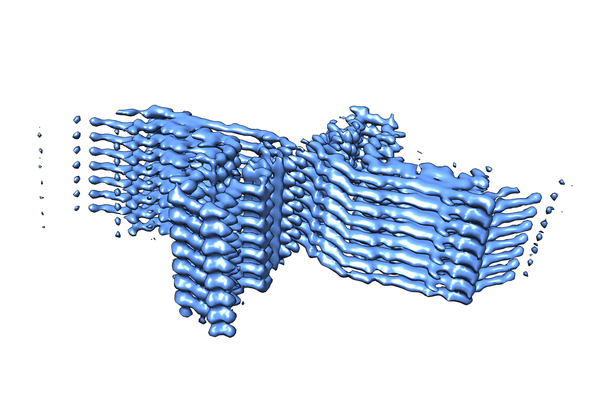
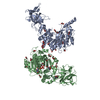
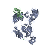
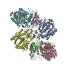
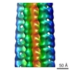
| タイトル | Two protofilament beta-2-microglobulin amyloid fibril | ||||||||||||||||||
 マップデータ マップデータ | Beta-2-microglobulin amyloid fibril - two protofilamets | ||||||||||||||||||
 試料 試料 |
| ||||||||||||||||||
| 機能・相同性 |  機能・相同性情報 機能・相同性情報positive regulation of ferrous iron binding / positive regulation of transferrin receptor binding / negative regulation of receptor binding / DAP12 interactions / positive regulation of receptor binding / early endosome lumen / Nef mediated downregulation of MHC class I complex cell surface expression / cellular response to iron ion / Endosomal/Vacuolar pathway / Antigen Presentation: Folding, assembly and peptide loading of class I MHC ...positive regulation of ferrous iron binding / positive regulation of transferrin receptor binding / negative regulation of receptor binding / DAP12 interactions / positive regulation of receptor binding / early endosome lumen / Nef mediated downregulation of MHC class I complex cell surface expression / cellular response to iron ion / Endosomal/Vacuolar pathway / Antigen Presentation: Folding, assembly and peptide loading of class I MHC / cellular response to iron(III) ion / antigen processing and presentation of exogenous protein antigen via MHC class Ib, TAP-dependent / negative regulation of forebrain neuron differentiation / regulation of erythrocyte differentiation / peptide antigen assembly with MHC class I protein complex / ER to Golgi transport vesicle membrane / regulation of iron ion transport / response to molecule of bacterial origin / MHC class I peptide loading complex / HFE-transferrin receptor complex / T cell mediated cytotoxicity / positive regulation of T cell cytokine production / antigen processing and presentation of endogenous peptide antigen via MHC class I / sensory perception of smell / MHC class I protein complex / negative regulation of neurogenesis / positive regulation of receptor-mediated endocytosis / peptide antigen assembly with MHC class II protein complex / multicellular organismal-level iron ion homeostasis / MHC class II protein complex / cellular response to nicotine / specific granule lumen / positive regulation of T cell mediated cytotoxicity / positive regulation of cellular senescence / recycling endosome membrane / phagocytic vesicle membrane / peptide antigen binding / antigen processing and presentation of exogenous peptide antigen via MHC class II / negative regulation of epithelial cell proliferation / Immunoregulatory interactions between a Lymphoid and a non-Lymphoid cell / positive regulation of immune response / Interferon gamma signaling / positive regulation of T cell activation / Modulation by Mtb of host immune system / negative regulation of neuron projection development / positive regulation of protein binding / tertiary granule lumen / DAP12 signaling / MHC class II protein complex binding / late endosome membrane / iron ion transport / ER-Phagosome pathway / T cell differentiation in thymus / early endosome membrane / protein refolding / protein homotetramerization / intracellular iron ion homeostasis / amyloid fibril formation / learning or memory / Amyloid fiber formation / endoplasmic reticulum lumen / lysosomal membrane / Golgi membrane / external side of plasma membrane / focal adhesion / Neutrophil degranulation / SARS-CoV-2 activates/modulates innate and adaptive immune responses / structural molecule activity / Golgi apparatus / endoplasmic reticulum / protein homodimerization activity / extracellular space / extracellular exosome / extracellular region / identical protein binding / membrane / plasma membrane / cytosol 類似検索 - 分子機能 | ||||||||||||||||||
| 生物種 |  Homo sapiens (ヒト) Homo sapiens (ヒト) | ||||||||||||||||||
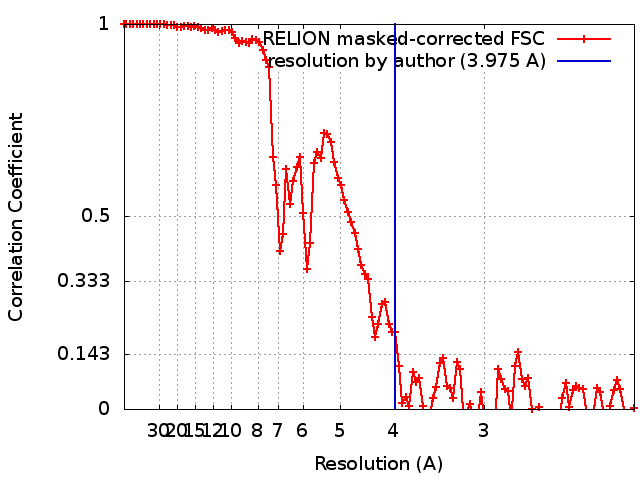
| 手法 | らせん対称体再構成法 / クライオ電子顕微鏡法 / 解像度: 3.975 Å | ||||||||||||||||||
 データ登録者 データ登録者 | Iadanza MG / Ranson NA | ||||||||||||||||||
| 資金援助 |  英国, 英国,  米国, 5件 米国, 5件
| ||||||||||||||||||
 引用 引用 | ジャーナル: Nat Commun / 年: 2018 タイトル: The structure of a β-microglobulin fibril suggests a molecular basis for its amyloid polymorphism. 著者: Matthew G Iadanza / Robert Silvers / Joshua Boardman / Hugh I Smith / Theodoros K Karamanos / Galia T Debelouchina / Yongchao Su / Robert G Griffin / Neil A Ranson / Sheena E Radford / 要旨: All amyloid fibrils contain a cross-β fold. How this structure differs in fibrils formed from proteins associated with different diseases remains unclear. Here, we combine cryo-EM and MAS-NMR to ...All amyloid fibrils contain a cross-β fold. How this structure differs in fibrils formed from proteins associated with different diseases remains unclear. Here, we combine cryo-EM and MAS-NMR to determine the structure of an amyloid fibril formed in vitro from β-microglobulin (βm), the culprit protein of dialysis-related amyloidosis. The fibril is composed of two identical protofilaments assembled from subunits that do not share βm's native tertiary fold, but are formed from similar β-strands. The fibrils share motifs with other amyloid fibrils, but also contain unique features including π-stacking interactions perpendicular to the fibril axis and an intramolecular disulfide that stabilises the subunit fold. We also describe a structural model for a second fibril morphology and show that it is built from the same subunit fold. The results provide insights into the mechanisms of fibril formation and the commonalities and differences within the amyloid fold in different protein sequences. | ||||||||||||||||||
| 履歴 |
|
- 構造の表示
構造の表示
| 構造ビューア | EMマップ: SurfViewMolmilJmol/JSmol |
|---|---|
| 添付画像 |
- ダウンロードとリンク
-EMDBアーカイブ
| マップデータ | emd_0014.map.gz | 5.3 MB | EMDBマップデータ形式 | |
|---|---|---|---|---|
| ヘッダ (付随情報) | emd-0014-v30.xmlemd-0014.xml | 15.3 KB 15.3 KB | 表示 表示 | EMDBヘッダ |
| FSC (解像度算出) | emd_0014_fsc.xml | 10.7 KB | 表示 | FSCデータファイル |
| 画像 |  emd_0014.png emd_0014.png | 163.4 KB | ||
| アーカイブディレクトリ |  http://ftp.pdbj.org/pub/emdb/structures/EMD-0014ftp://ftp.pdbj.org/pub/emdb/structures/EMD-0014 http://ftp.pdbj.org/pub/emdb/structures/EMD-0014ftp://ftp.pdbj.org/pub/emdb/structures/EMD-0014 | HTTPS FTP |
-検証レポート
| 文書・要旨 | emd_0014_validation.pdf.gz | 326.9 KB | 表示 | EMDB検証レポート |
|---|---|---|---|---|
| 文書・詳細版 | emd_0014_full_validation.pdf.gz | 326.5 KB | 表示 | |
| XML形式データ | emd_0014_validation.xml.gz | 11.9 KB | 表示 | |
| CIF形式データ | emd_0014_validation.cif.gz | 15.8 KB | 表示 | |
| アーカイブディレクトリ | https://ftp.pdbj.org/pub/emdb/validation_reports/EMD-0014ftp://ftp.pdbj.org/pub/emdb/validation_reports/EMD-0014 | HTTPS FTP |
-関連構造データ
| 関連構造データ |  6gk3MC 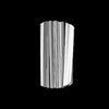 0021C M: このマップから作成された原子モデル C: 同じ文献を引用 ( |
|---|---|
| 類似構造データ | |
| 電子顕微鏡画像生データ | EMPIAR-10207 (タイトル: Beta-2-microglobulin fibrils with multiple polymorphs formed at pH 2 Data size: 294.3 Data #1: Aligned, dose-weighted micrographs [micrographs - single frame]) |


-リンク
| EMDBのページ | EMDB (EBI/PDBe) / EMDataResource |
|---|---|
| 「今月の分子」の関連する項目 |


-マップ
| ファイル | ダウンロード / ファイル: emd_0014.map.gz / 形式: CCP4 / 大きさ: 103 MB / タイプ: IMAGE STORED AS FLOATING POINT NUMBER (4 BYTES) | ||||||||||||||||||||||||||||||||||||
|---|---|---|---|---|---|---|---|---|---|---|---|---|---|---|---|---|---|---|---|---|---|---|---|---|---|---|---|---|---|---|---|---|---|---|---|---|---|
| 注釈 | Beta-2-microglobulin amyloid fibril - two protofilamets | ||||||||||||||||||||||||||||||||||||
| 投影像・断面図 | 画像のコントロール
画像は Spider により作成 | ||||||||||||||||||||||||||||||||||||
| ボクセルのサイズ | X=Y=Z: 1.06 Å | ||||||||||||||||||||||||||||||||||||
| 密度 |
| ||||||||||||||||||||||||||||||||||||
| 対称性 | 空間群: 1 | ||||||||||||||||||||||||||||||||||||
| 詳細 | EMDB XML:
|
 Z (Sec.)
 Y (Row.)
 X (Col.)


-添付データ
- 試料の構成要素
-全体 : two protofilament beta-2-microglobulin amyloid fibril
| 全体 | 名称: two protofilament beta-2-microglobulin amyloid fibril |
|---|---|
| 要素 |
|
-超分子 #1: two protofilament beta-2-microglobulin amyloid fibril
| 超分子 | 名称: two protofilament beta-2-microglobulin amyloid fibril タイプ: complex / ID: 1 / 親要素: 0 / 含まれる分子: all |
|---|---|
| 由来(天然) | 生物種: Homo sapiens (ヒト) |
| 組換発現 | 生物種:  |
| 分子量 | 理論値: 53.3 kDa/nm |
-分子 #1: Beta-2-microglobulin
| 分子 | 名称: Beta-2-microglobulin / タイプ: protein_or_peptide / ID: 1 / コピー数: 8 / 光学異性体: LEVO |
|---|---|
| 由来(天然) | 生物種: Homo sapiens (ヒト) |
| 分子量 | 理論値: 7.635446 KDa |
| 組換発現 | 生物種: |
| 配列 | 文字列: FLNCYVSGFH PSDIEVDLLK NGERIEKVEH SDLSFSKDWS FYLLYYTEFT PTEKDEYACR VNHV |
-実験情報
-構造解析
| 手法 | クライオ電子顕微鏡法 |
|---|---|
 解析 解析 | らせん対称体再構成法 |
| 試料の集合状態 | filament |
-試料調製
| 濃度 | 0.025 mg/mL | ||||||||||||
|---|---|---|---|---|---|---|---|---|---|---|---|---|---|
| 緩衝液 | pH: 2.5 構成要素:
| ||||||||||||
| グリッド | モデル: Quantifoil R3.5/1 / 材質: COPPER / メッシュ: 400 / 支持フィルム - 材質: CARBON / 支持フィルム - トポロジー: HOLEY / 前処理 - タイプ: PLASMA CLEANING | ||||||||||||
| 凍結 | 凍結剤: ETHANE / チャンバー内湿度: 80 % / チャンバー内温度: 281.15 K / 装置: FEI VITROBOT MARK II | ||||||||||||
| 詳細 | Quiescent growth at 0.25 mg/ml for 5 weeks, diluted 10x with buffer |
- 電子顕微鏡法
| 顕微鏡 | FEI TITAN KRIOS |
|---|---|
| 撮影 | フィルム・検出器のモデル: GATAN K2 SUMMIT (4k x 4k) 検出モード: COUNTING / デジタル化 - 画像ごとのフレーム数: 3-40 / 撮影したグリッド数: 1 / 実像数: 5549 / 平均露光時間: 10.0 sec. / 平均電子線量: 38.5 e/Å2 |
| 電子線 | 加速電圧: 300 kV / 電子線源:  FIELD EMISSION GUN FIELD EMISSION GUN |
| 電子光学系 | C2レンズ絞り径: 100.0 µm / 照射モード: FLOOD BEAM / 撮影モード: BRIGHT FIELD / Cs: 2.7 mm 最大 デフォーカス(公称値): 0.0032500000000000003 µm 最小 デフォーカス(公称値): 0.00175 µm / 倍率(公称値): 130000 |
| 試料ステージ | 試料ホルダーモデル: FEI TITAN KRIOS AUTOGRID HOLDER ホルダー冷却材: NITROGEN |
| 実験機器 |  モデル: Titan Krios / 画像提供: FEI Company |
+画像解析
-原子モデル構築 1
| 精密化 | 空間: REAL / プロトコル: OTHER |
|---|---|
| 得られたモデル | PDB-6gk3: |
