 ムービー
ムービー コントローラー


+ データを開く

- 基本情報
基本情報
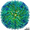
| 登録情報 | データベース: EMDB / ID: EMD-22972 | |||||||||||||||||||||
|---|---|---|---|---|---|---|---|---|---|---|---|---|---|---|---|---|---|---|---|---|---|---|
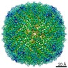
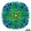
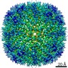
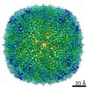
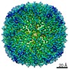
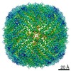
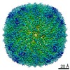
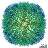
| タイトル | Cryo-EM structure of heavy chain mouse apoferritin | |||||||||||||||||||||
 マップデータ マップデータ | raw unsharpened map | |||||||||||||||||||||
 試料 試料 |
| |||||||||||||||||||||
 キーワード キーワード | protein / OXIDOREDUCTASE | |||||||||||||||||||||
| 機能・相同性 |  機能・相同性情報 機能・相同性情報Iron uptake and transport / Golgi Associated Vesicle Biogenesis / negative regulation of ferroptosis / iron ion sequestering activity / autolysosome / ferroxidase / intracellular sequestering of iron ion / ferroxidase activity / negative regulation of fibroblast proliferation / endocytic vesicle lumen ...Iron uptake and transport / Golgi Associated Vesicle Biogenesis / negative regulation of ferroptosis / iron ion sequestering activity / autolysosome / ferroxidase / intracellular sequestering of iron ion / ferroxidase activity / negative regulation of fibroblast proliferation / endocytic vesicle lumen / Neutrophil degranulation / ferric iron binding / ferrous iron binding / iron ion transport / iron ion binding / immune response / negative regulation of cell population proliferation / mitochondrion / extracellular region / identical protein binding / membrane / cytoplasm / cytosol 類似検索 - 分子機能 | |||||||||||||||||||||
| 生物種 |  | |||||||||||||||||||||
| 手法 | 単粒子再構成法 / クライオ電子顕微鏡法 / 解像度: 1.655 Å | |||||||||||||||||||||
 データ登録者 データ登録者 | Sun M / Azumaya C | |||||||||||||||||||||
| 資金援助 |  米国, 6件 米国, 6件
| |||||||||||||||||||||
 引用 引用 | ジャーナル: J Struct Biol / 年: 2021 タイトル: Practical considerations for using K3 cameras in CDS mode for high-resolution and high-throughput single particle cryo-EM. 著者: Ming Sun / Caleigh M Azumaya / Eric Tse / David P Bulkley / Matthew B Harrington / Glenn Gilbert / Adam Frost / Daniel Southworth / Kliment A Verba / Yifan Cheng / David A Agard / 要旨: Detector technology plays a pivotal role in high-resolution and high-throughput cryo-EM structure determination. Compared with the first-generation, single-electron counting direct detection camera ...Detector technology plays a pivotal role in high-resolution and high-throughput cryo-EM structure determination. Compared with the first-generation, single-electron counting direct detection camera (Gatan K2), the latest K3 camera is faster, larger, and now offers a correlated-double sampling mode (CDS). Importantly this results in a higher DQE and improved throughput compared to its predecessor. In this study, we focused on optimizing camera data collection parameters for daily use within a cryo-EM facility and explored the balance between throughput and resolution. In total, eight data sets of murine heavy-chain apoferritin were collected at different dose rates and magnifications, using 9-hole image shift data collection strategies. The performance of the camera was characterized by the quality of the resultant 3D reconstructions. Our results demonstrated that the Gatan K3 operating in CDS mode outperformed standard (nonCDS) mode in terms of reconstruction resolution in all tested conditions with 8 electrons per pixel per second being the optimal dose rate. At low magnification (64kx) we were able to achieve reconstruction resolutions of 149% of the physical Nyquist limit (1.8 Å with a 1.346 Å physical pixel size). Low magnification allows more particles to be collected per image, aiding analysis of heterogeneous samples requiring large data sets. At moderate magnification (105kx, 0.834 Å physical pixel size) we achieved a resolution of 1.65 Å within 8-h of data collection, a condition optimal for achieving high-resolution on well behaved samples. Our results also show that for an optimal sample like apoferritin, one can achieve better than 2.5 Å resolution with 5 min of data collection. Together, our studies validate the most efficient ways of imaging protein complexes using the K3 direct detector and will greatly benefit the cryo-EM community. | |||||||||||||||||||||
| 履歴 |
|
- 構造の表示
構造の表示
| ムービー |
ムービービューア |
|---|---|
| 構造ビューア | EMマップ: SurfViewMolmilJmol/JSmol |
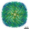
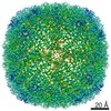
| 添付画像 |

- ダウンロードとリンク
-EMDBアーカイブ
| マップデータ | emd_22972.map.gz | 455.6 MB | EMDBマップデータ形式 | |
|---|---|---|---|---|
| ヘッダ (付随情報) | emd-22972-v30.xmlemd-22972.xml | 17.4 KB 17.4 KB | 表示 表示 | EMDBヘッダ |
| FSC (解像度算出) | emd_22972_fsc.xml | 18.1 KB | 表示 | FSCデータファイル |
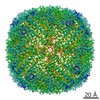
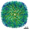
| 画像 | 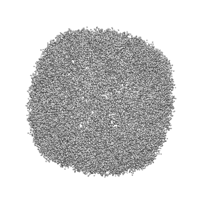 emd_22972.png emd_22972.png | 68.2 KB | ||
| Filedesc metadata | emd-22972.cif.gz | 5.6 KB | ||
| その他 | emd_22972_additional_1.map.gzemd_22972_additional_2.map.gz | 76.9 MB 72.2 MB | ||
| アーカイブディレクトリ |  http://ftp.pdbj.org/pub/emdb/structures/EMD-22972ftp://ftp.pdbj.org/pub/emdb/structures/EMD-22972 http://ftp.pdbj.org/pub/emdb/structures/EMD-22972ftp://ftp.pdbj.org/pub/emdb/structures/EMD-22972 | HTTPS FTP |
-検証レポート
| 文書・要旨 | emd_22972_validation.pdf.gz | 452.8 KB | 表示 | EMDB検証レポート |
|---|---|---|---|---|
| 文書・詳細版 | emd_22972_full_validation.pdf.gz | 452.4 KB | 表示 | |
| XML形式データ | emd_22972_validation.xml.gz | 15.9 KB | 表示 | |
| CIF形式データ | emd_22972_validation.cif.gz | 21.9 KB | 表示 | |
| アーカイブディレクトリ | https://ftp.pdbj.org/pub/emdb/validation_reports/EMD-22972ftp://ftp.pdbj.org/pub/emdb/validation_reports/EMD-22972 | HTTPS FTP |
-関連構造データ
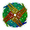
| 関連構造データ | 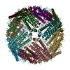 7kodMC M: このマップから作成された原子モデル C: 同じ文献を引用 ( |
|---|---|
| 類似構造データ | |
| 電子顕微鏡画像生データ | EMPIAR-10588 (タイトル: Cryo-EM reconstruction of heavy chain mouse apoferritin, 8 datasets testing dose rate and correlative double sampling mode for the Gatan K3 direct electron detector. [1143 multi-frame ...タイトル: Cryo-EM reconstruction of heavy chain mouse apoferritin, 8 datasets testing dose rate and correlative double sampling mode for the Gatan K3 direct electron detector. [1143 multi-frame micrographs composed of 120 frames in LZW compressed TIFF format Data size: 8.5 TB Data #1: apoferritin multi-frame raw micrographs non gain normalized frames in LZW compressed TIFF format collected at 8 e/p/s in CDS mode from Gatan K3 [micrographs - multiframe] Data #2: apoferritin multi-frame raw micrographs non gain normalized frames in LZW compressed TIFF format collected at 8 e/p/s in standard mode from Gatan K3 [micrographs - multiframe] Data #3: apoferritin multi-frame raw micrographs non gain normalized frames in LZW compressed TIFF format collected at 4 e/p/s in CDS mode from Gatan K3 [micrographs - multiframe] Data #4: apoferritin multi-frame raw micrographs non gain normalized frames in LZW compressed TIFF format collected at 4 e/p/s in CDS mode from Gatan K3 [micrographs - multiframe] Data #5: apoferritin multi-frame raw micrographs non gain normalized frames in LZW compressed TIFF format collected at 8 e/p/s in CDS mode from Gatan K3 at lower magnification [micrographs - multiframe] Data #6: apoferritin multi-frame raw micrographs non gain normalized frames in LZW compressed TIFF format collected at 8 e/p/s in standard mode from Gatan K3 at lower magnification [micrographs - multiframe] Data #7: apoferritin multi-frame raw micrographs non gain normalized frames in LZW compressed TIFF format collected at 16 e/p/s in CDS mode from Gatan K3 at lower magnification [micrographs - multiframe] Data #8: apoferritin multi-frame raw micrographs non gain normalized frames in LZW compressed TIFF format collected at 16 e/p/s in standard mode from Gatan K3 at lower magnification [micrographs - multiframe]) |


-リンク
| EMDBのページ | EMDB (EBI/PDBe) / EMDataResource |
|---|---|
| 「今月の分子」の関連する項目 |


-マップ
| ファイル | ダウンロード / ファイル: emd_22972.map.gz / 形式: CCP4 / 大きさ: 512 MB / タイプ: IMAGE STORED AS FLOATING POINT NUMBER (4 BYTES) | ||||||||||||||||||||||||||||||||||||||||||||||||||||||||||||||||||||
|---|---|---|---|---|---|---|---|---|---|---|---|---|---|---|---|---|---|---|---|---|---|---|---|---|---|---|---|---|---|---|---|---|---|---|---|---|---|---|---|---|---|---|---|---|---|---|---|---|---|---|---|---|---|---|---|---|---|---|---|---|---|---|---|---|---|---|---|---|---|
| 注釈 | raw unsharpened map | ||||||||||||||||||||||||||||||||||||||||||||||||||||||||||||||||||||
| ボクセルのサイズ | X=Y=Z: 0.417 Å | ||||||||||||||||||||||||||||||||||||||||||||||||||||||||||||||||||||
| 密度 |
| ||||||||||||||||||||||||||||||||||||||||||||||||||||||||||||||||||||
| 対称性 | 空間群: 1 | ||||||||||||||||||||||||||||||||||||||||||||||||||||||||||||||||||||
| 詳細 | EMDB XML:
CCP4マップ ヘッダ情報:
| ||||||||||||||||||||||||||||||||||||||||||||||||||||||||||||||||||||
-添付データ
-追加マップ: filtered and sharpened map (b-factor, ~ -66)
| ファイル | emd_22972_additional_1.map | ||||||||||||
|---|---|---|---|---|---|---|---|---|---|---|---|---|---|
| 注釈 | filtered and sharpened map (b-factor, ~ -66) | ||||||||||||
| 投影像・断面図 |
| ||||||||||||
| 密度ヒストグラム |
 Z
Z Y
Y X
X


-追加マップ: filtered and sharpened map (b-factor, ~ -25)
| ファイル | emd_22972_additional_2.map | ||||||||||||
|---|---|---|---|---|---|---|---|---|---|---|---|---|---|
| 注釈 | filtered and sharpened map (b-factor, ~ -25) | ||||||||||||
| 投影像・断面図 |
| ||||||||||||
| 密度ヒストグラム |


- 試料の構成要素
-全体 : Heavy chain mouse apoferritin
| 全体 | 名称: Heavy chain mouse apoferritin |
|---|---|
| 要素 |
|
-超分子 #1: Heavy chain mouse apoferritin
| 超分子 | 名称: Heavy chain mouse apoferritin / タイプ: complex / ID: 1 / 親要素: 0 / 含まれる分子: #1 |
|---|---|
| 由来(天然) | 生物種: |
-分子 #1: Ferritin heavy chain
| 分子 | 名称: Ferritin heavy chain / タイプ: protein_or_peptide / ID: 1 / コピー数: 24 / 光学異性体: LEVO / EC番号: ferroxidase |
|---|---|
| 由来(天然) | 生物種: |
| 分子量 | 理論値: 21.097631 KDa |
| 組換発現 | 生物種:  |
| 配列 | 文字列: MTTASPSQVR QNYHQDAEAA INRQINLELY ASYVYLSMSC YFDRDDVALK NFAKYFLHQS HEEREHAEKL MKLQNQRGGR IFLQDIKKP DRDDWESGLN AMECALHLEK SVNQSLLELH KLATDKNDPH LCDFIETYYL SEQVKSIKEL GDHVTNLRKM G APEAGMAE YLFDKHTLGH GDES UniProtKB: Ferritin heavy chain |
-分子 #2: water
| 分子 | 名称: water / タイプ: ligand / ID: 2 / コピー数: 1727 / 式: HOH |
|---|---|
| 分子量 | 理論値: 18.015 Da |
| Chemical component information |  ChemComp-HOH: |
-実験情報
-構造解析
| 手法 | クライオ電子顕微鏡法 |
|---|---|
 解析 解析 | 単粒子再構成法 |
| 試料の集合状態 | particle |
-試料調製
| 緩衝液 | pH: 7.5 |
|---|---|
| グリッド | モデル: Quantifoil / 材質: COPPER / メッシュ: 300 / 前処理 - タイプ: GLOW DISCHARGE |
| 凍結 | 凍結剤: ETHANE |
- 電子顕微鏡法
| 顕微鏡 | FEI TITAN KRIOS |
|---|---|
| 撮影 | フィルム・検出器のモデル: GATAN K3 BIOQUANTUM (6k x 4k) 撮影したグリッド数: 1 / 平均露光時間: 6.0 sec. / 平均電子線量: 69.0 e/Å2 / 詳細: 9-hole image-shift data collection strategy |
| 電子線 | 加速電圧: 300 kV / 電子線源:  FIELD EMISSION GUN FIELD EMISSION GUN |
| 電子光学系 | 照射モード: FLOOD BEAM / 撮影モード: BRIGHT FIELD |
| 試料ステージ | 試料ホルダーモデル: FEI TITAN KRIOS AUTOGRID HOLDER |
| 実験機器 |  モデル: Titan Krios / 画像提供: FEI Company |


