 Movie
Movie Controller

[English] 日本語
 Yorodumi
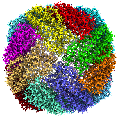
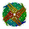
- EMDB-22658: Apoferritin structure at 1.36 angstrom resolution determined from... -
+ Open data

- Basic information
| Entry | Database: EMDB / ID: EMD-22658 | ||||||||||||
|---|---|---|---|---|---|---|---|---|---|---|---|---|---|
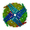
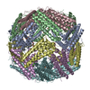
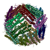
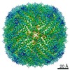
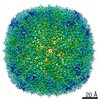
| Title | Apoferritin structure at 1.36 angstrom resolution determined from a 300 kV Titan Krios G3i electron microscope with Falcon4 detector | ||||||||||||
 Map data Map data | |||||||||||||
 Sample Sample |
| ||||||||||||
 Keywords Keywords | Apoferritin / cryo-EM / Atomic resolution / Falcon4 detector / OXIDOREDUCTASE | ||||||||||||
| Function / homology |  Function and homology information Function and homology informationiron ion sequestering activity / : / autolysosome / Scavenging by Class A Receptors / Golgi Associated Vesicle Biogenesis / ferroxidase / intracellular sequestering of iron ion / ferroxidase activity / negative regulation of fibroblast proliferation / ferric iron binding ...iron ion sequestering activity / : / autolysosome / Scavenging by Class A Receptors / Golgi Associated Vesicle Biogenesis / ferroxidase / intracellular sequestering of iron ion / ferroxidase activity / negative regulation of fibroblast proliferation / ferric iron binding / Iron uptake and transport / ferrous iron binding / tertiary granule lumen / iron ion transport / intracellular iron ion homeostasis / ficolin-1-rich granule lumen / iron ion binding / immune response / negative regulation of cell population proliferation / Neutrophil degranulation / extracellular exosome / extracellular region / identical protein binding / membrane / nucleus / cytoplasm / cytosol Similarity search - Function | ||||||||||||
| Biological species |  Homo sapiens (human) Homo sapiens (human) | ||||||||||||
| Method | single particle reconstruction / cryo EM / Resolution: 1.36 Å | ||||||||||||
 Authors Authors | Zhang K / Pintilie G | ||||||||||||
| Funding support |  United States, 3 items United States, 3 items
| ||||||||||||
 Citation Citation | Journal: Cell Res / Year: 2020 Title: Resolving individual atoms of protein complex by cryo-electron microscopy. Authors: Kaiming Zhang / Grigore D Pintilie / Shanshan Li / Michael F Schmid / Wah Chiu / | ||||||||||||
| History |
|
- Structure visualization
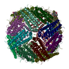
| Movie |
Movie viewer |
|---|---|
| Structure viewer | EM map: SurfViewMolmilJmol/JSmol |
| Supplemental images |
- Downloads & links
-EMDB archive
| Map data | emd_22658.map.gz | 124.5 MB | EMDB map data format | |
|---|---|---|---|---|
| Header (meta data) | emd-22658-v30.xmlemd-22658.xml | 12.9 KB 12.9 KB | Display Display | EMDB header |
| Images |  emd_22658.png emd_22658.png | 320.3 KB | ||
| Filedesc metadata | emd-22658.cif.gz | 5.6 KB | ||
| Archive directory |  http://ftp.pdbj.org/pub/emdb/structures/EMD-22658ftp://ftp.pdbj.org/pub/emdb/structures/EMD-22658 http://ftp.pdbj.org/pub/emdb/structures/EMD-22658ftp://ftp.pdbj.org/pub/emdb/structures/EMD-22658 | HTTPS FTP |
-Validation report
| Summary document | emd_22658_validation.pdf.gz | 779.8 KB | Display | EMDB validaton report |
|---|---|---|---|---|
| Full document | emd_22658_full_validation.pdf.gz | 779.3 KB | Display | |
| Data in XML | emd_22658_validation.xml.gz | 6.5 KB | Display | |
| Data in CIF | emd_22658_validation.cif.gz | 7.4 KB | Display | |
| Arichive directory | https://ftp.pdbj.org/pub/emdb/validation_reports/EMD-22658ftp://ftp.pdbj.org/pub/emdb/validation_reports/EMD-22658 | HTTPS FTP |
-Related structure data
| Related structure data |  7k3wMC  7k3vC  7rrpC C: citing same article ( M: atomic model generated by this map |
|---|---|
| Similar structure data | |
| EM raw data | EMPIAR-10635 (Title: Apoferritin structure at 1.36 angstrom resolution determined from a 300 kV Titan Krios G3i electron microscope with Falcon4 detector Data size: 9.4 TB Data #1: Apoferritin structure at 1.36 angstrom resolution determined from a 300 kV Titan Krios G3i electron microscope with Falcon4 detector [micrographs - multiframe]) |


-Links
| EMDB pages | EMDB (EBI/PDBe) / EMDataResource |
|---|---|
| Related items in Molecule of the Month |


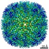
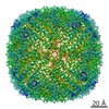
-Map
| File | Download / File: emd_22658.map.gz / Format: CCP4 / Size: 137.1 MB / Type: IMAGE STORED AS FLOATING POINT NUMBER (4 BYTES) | ||||||||||||||||||||||||||||||||||||||||||||||||||||||||||||||||||||
|---|---|---|---|---|---|---|---|---|---|---|---|---|---|---|---|---|---|---|---|---|---|---|---|---|---|---|---|---|---|---|---|---|---|---|---|---|---|---|---|---|---|---|---|---|---|---|---|---|---|---|---|---|---|---|---|---|---|---|---|---|---|---|---|---|---|---|---|---|---|
| Voxel size | X=Y=Z: 0.502 Å | ||||||||||||||||||||||||||||||||||||||||||||||||||||||||||||||||||||
| Density |
| ||||||||||||||||||||||||||||||||||||||||||||||||||||||||||||||||||||
| Symmetry | Space group: 1 | ||||||||||||||||||||||||||||||||||||||||||||||||||||||||||||||||||||
| Details | EMDB XML:
CCP4 map header:
| ||||||||||||||||||||||||||||||||||||||||||||||||||||||||||||||||||||
-Supplemental data
- Sample components
-Entire : Apoferritin
| Entire | Name: Apoferritin |
|---|---|
| Components |
|
-Supramolecule #1: Apoferritin
| Supramolecule | Name: Apoferritin / type: complex / ID: 1 / Parent: 0 / Macromolecule list: #1 |
|---|---|
| Source (natural) | Organism: Homo sapiens (human) |
| Molecular weight | Theoretical: 480 KDa |
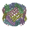
-Macromolecule #1: Ferritin heavy chain
| Macromolecule | Name: Ferritin heavy chain / type: protein_or_peptide / ID: 1 / Number of copies: 24 / Enantiomer: LEVO / EC number: ferroxidase |
|---|---|
| Source (natural) | Organism: Homo sapiens (human) |
| Molecular weight | Theoretical: 20.116547 KDa |
| Recombinant expression | Organism:  |
| Sequence | String: TSQVRQNYHQ DSEAAINRQI NLELYASYVY LSMSYYFDRD DVALKNFAKY FLHQSHEERE HAEKLMKLQN QRGGRIFLQD IKKPDCDDW ESGLNAMECA LHLEKNVNQS LLELHKLATD KNDPHLCDFI ETHYLNEQVK AIKELGDHVT NLRKMGAPES G LAEYLFDK HTLG UniProtKB: Ferritin heavy chain |
-Macromolecule #2: ZINC ION
| Macromolecule | Name: ZINC ION / type: ligand / ID: 2 / Number of copies: 270 / Formula: ZN |
|---|---|
| Molecular weight | Theoretical: 65.409 Da |
-Macromolecule #3: SODIUM ION
| Macromolecule | Name: SODIUM ION / type: ligand / ID: 3 / Number of copies: 112 |
|---|---|
| Molecular weight | Theoretical: 22.99 Da |
-Macromolecule #4: water
| Macromolecule | Name: water / type: ligand / ID: 4 / Number of copies: 2680 / Formula: HOH |
|---|---|
| Molecular weight | Theoretical: 18.015 Da |
| Chemical component information |  ChemComp-HOH: |
-Experimental details
-Structure determination
| Method | cryo EM |
|---|---|
 Processing Processing | single particle reconstruction |
| Aggregation state | particle |
-Sample preparation
| Concentration | 0.2 mg/mL |
|---|---|
| Buffer | pH: 8 / Details: 50 mM Tris-HCl (pH 8.0), 150 mM NaCl |
| Grid | Model: Quantifoil R2/1 / Material: COPPER / Mesh: 200 / Support film - Material: CARBON / Support film - topology: CONTINUOUS / Support film - Film thickness: 0.2 / Pretreatment - Type: GLOW DISCHARGE / Pretreatment - Time: 20 sec. |
| Vitrification | Cryogen name: ETHANE |
- Electron microscopy
| Microscope | FEI TITAN KRIOS |
|---|---|
| Image recording | Film or detector model: FEI FALCON IV (4k x 4k) / Average electron dose: 40.0 e/Å2 |
| Electron beam | Acceleration voltage: 300 kV / Electron source:  FIELD EMISSION GUN FIELD EMISSION GUN |
| Electron optics | C2 aperture diameter: 70.0 µm / Illumination mode: FLOOD BEAM / Imaging mode: BRIGHT FIELD / Cs: 2.7 mm |
| Sample stage | Specimen holder model: FEI TITAN KRIOS AUTOGRID HOLDER / Cooling holder cryogen: NITROGEN |
| Experimental equipment |  Model: Titan Krios / Image courtesy: FEI Company |
+Image processing
-Atomic model buiding 1
| Initial model | PDB ID: Chain - Chain ID: A / Chain - Source name: PDB / Chain - Initial model type: experimental model |
|---|---|
| Refinement | Protocol: OTHER / Target criteria: CC |
| Output model | PDB-7k3w: |

