National Institutes of Health/National Cancer Institute (NIH/NCI)
CA221289
United States
National Institutes of Health/National Heart, Lung, and Blood Institute (NIH/NHLBI)
HL122416
United States
National Institutes of Health/National Heart, Lung, and Blood Institute (NIH/NHLBI)
HL071818
United States
Citation
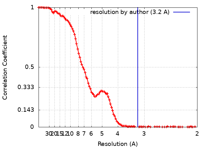
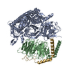
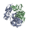
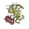
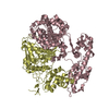
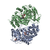
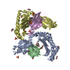
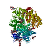
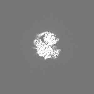
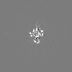
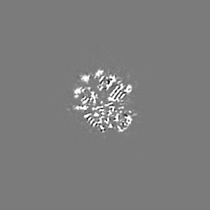
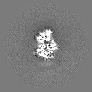
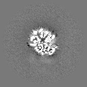
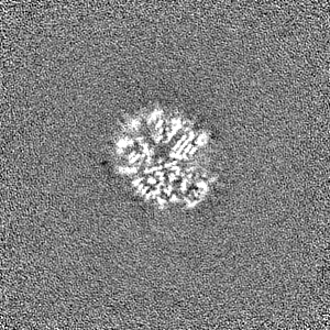
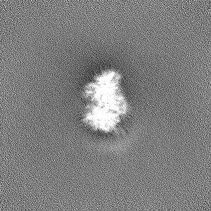
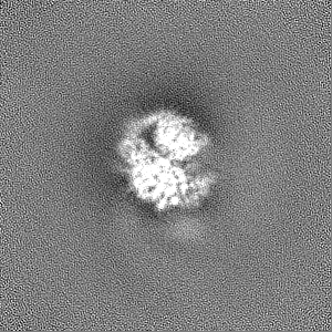
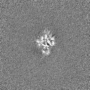
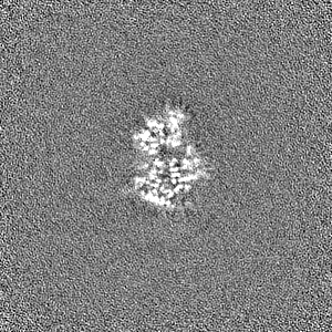
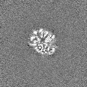
Journal: Sci Adv / Year: 2019 Title: Cryo-electron microscopy structure and analysis of the P-Rex1-Gβγ signaling scaffold. Authors: Jennifer N Cash / Sarah Urata / Sheng Li / Sandeep K Ravala / Larisa V Avramova / Michael D Shost / J Silvio Gutkind / John J G Tesmer / Michael A Cianfrocco / Abstract: PIP-dependent Rac exchanger 1 (P-Rex1) is activated downstream of G protein-coupled receptors to promote neutrophil migration and metastasis. The structure of more than half of the enzyme and its ...PIP-dependent Rac exchanger 1 (P-Rex1) is activated downstream of G protein-coupled receptors to promote neutrophil migration and metastasis. The structure of more than half of the enzyme and its regulatory G protein binding site are unknown. Our 3.2 Å cryo-EM structure of the P-Rex1-Gβγ complex reveals that the carboxyl-terminal half of P-Rex1 adopts a complex fold most similar to those of phosphoinositide phosphatases. Although catalytically inert, the domain coalesces with a DEP domain and two PDZ domains to form an extensive docking site for Gβγ. Hydrogen-deuterium exchange mass spectrometry suggests that Gβγ binding induces allosteric changes in P-Rex1, but functional assays indicate that membrane localization is also required for full activation. Thus, a multidomain assembly is key to the regulation of P-Rex1 by Gβγ and the formation of a membrane-localized scaffold optimized for recruitment of other signaling proteins such as PKA and PTEN.
History
Deposition
Jun 18, 2019
-
Header (metadata) release
Jul 3, 2019
-
Map release
Oct 23, 2019
-
Update
Mar 20, 2024
-
Current status
Mar 20, 2024
Processing site: RCSB / Status: Released
-
Structure visualization
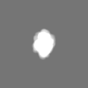
Movie
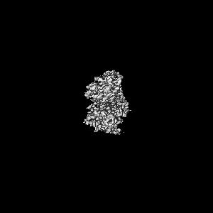
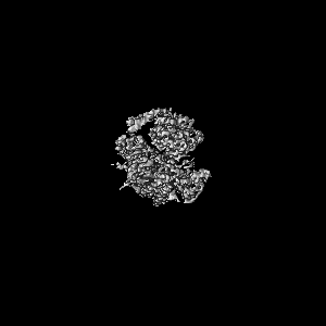
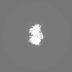
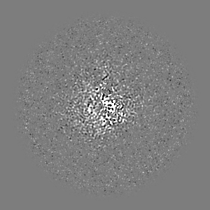
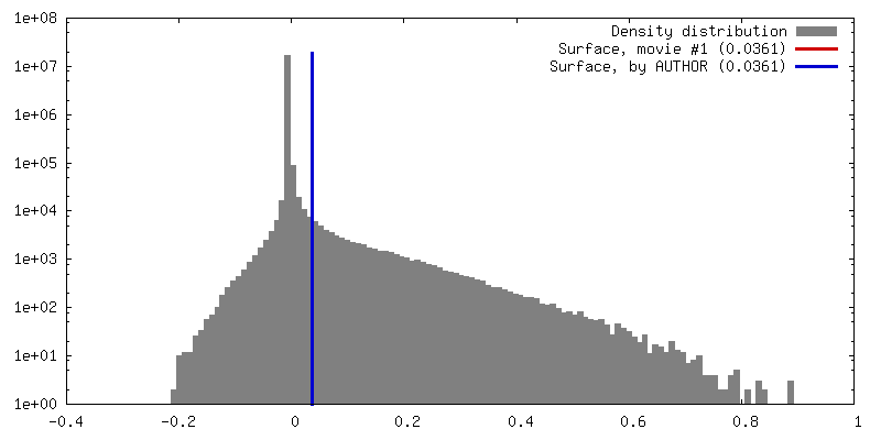
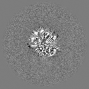
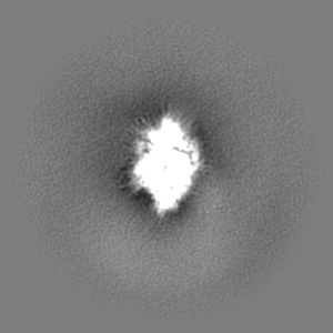
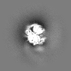
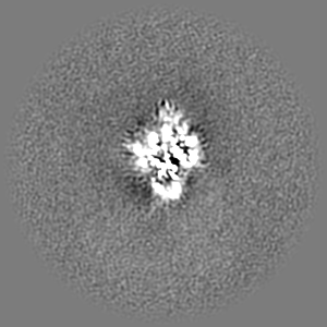
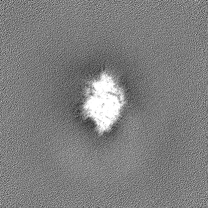
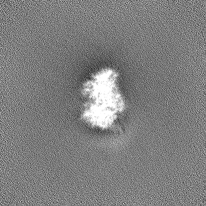
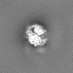
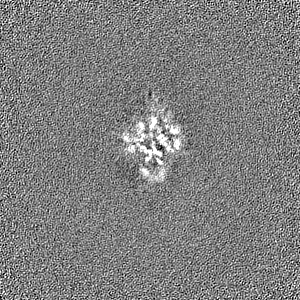
Surface view with section colored by density value
EMPIAR-10285 (Title: Cryo-electron microscopy structure of the P-Rex1–G-beta-gamma signaling scaffold Data size: 3.0 TB Data #1: Movie files (.tif) for P-Rex1-Gbg [micrographs - multiframe] Data #2: Micrograph files (.mrc) & CTF log files for P-Rex1-Gbg [micrographs - single frame] Data #3: Extracted particles from Warp for P-Rex1-Gbg [picked particles - single frame - processed])
chain_id: A, residue_range: 622-706, source_name: PDB, initial_model_type: experimental model
Refinement
Space: REAL / Protocol: AB INITIO MODEL / Overall B value: 83
Output model
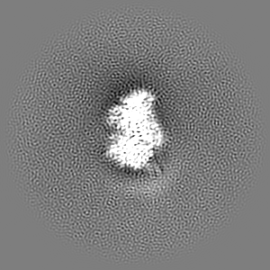
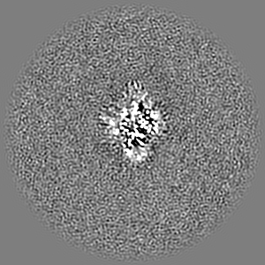
PDB-6pcv: Single Particle Reconstruction of Phosphatidylinositol (3,4,5) trisphosphate-dependent Rac exchanger 1 bound to G protein beta gamma subunits
+
About Yorodumi
-
News
-
Feb 9, 2022. New format data for meta-information of EMDB entries
New format data for meta-information of EMDB entries
Version 3 of the EMDB header file is now the official format.
The previous official version 1.9 will be removed from the archive.
In the structure databanks used in Yorodumi, some data are registered as the other names, "COVID-19 virus" and "2019-nCoV". Here are the details of the virus and the list of structure data.
Jan 31, 2019. EMDB accession codes are about to change! (news from PDBe EMDB page)
EMDB accession codes are about to change! (news from PDBe EMDB page)
The allocation of 4 digits for EMDB accession codes will soon come to an end. Whilst these codes will remain in use, new EMDB accession codes will include an additional digit and will expand incrementally as the available range of codes is exhausted. The current 4-digit format prefixed with “EMD-” (i.e. EMD-XXXX) will advance to a 5-digit format (i.e. EMD-XXXXX), and so on. It is currently estimated that the 4-digit codes will be depleted around Spring 2019, at which point the 5-digit format will come into force.
The EM Navigator/Yorodumi systems omit the EMD- prefix.
Related info.:Q: What is EMD? / ID/Accession-code notation in Yorodumi/EM Navigator
Yorodumi is a browser for structure data from EMDB, PDB, SASBDB, etc.
This page is also the successor to EM Navigator detail page, and also detail information page/front-end page for Omokage search.
The word "yorodu" (or yorozu) is an old Japanese word meaning "ten thousand". "mi" (miru) is to see.
Related info.:EMDB / PDB / SASBDB / Comparison of 3 databanks / Yorodumi Search / Aug 31, 2016. New EM Navigator & Yorodumi / Yorodumi Papers / Jmol/JSmol / Function and homology information / Changes in new EM Navigator and Yorodumi
 Movie
Movie Controller

 Yorodumi
 Open data
 Basic information
 Map data
 Sample
 Keywords
 Function and homology information
 Homo sapiens (human) /
 Authors
 United States, 3 items
  Citation
 Structure visualization
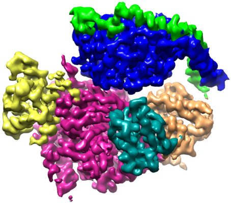
 Downloads & links
 emd_20308.png
 http://ftp.pdbj.org/pub/emdb/structures/EMD-20308


 Z (Sec.)
 Y (Row.)
 X (Col.)


 Sample components
 Trichoplusia ni (cabbage looper)
 Processing
 Electron microscopy
 FIELD EMISSION GUN