 Movie
Movie Controller


+ Open data

- Basic information
| Entry |  | ||||||||||||||||||
|---|---|---|---|---|---|---|---|---|---|---|---|---|---|---|---|---|---|---|---|
| Title | Cryo-EM structure of the 6 hpf zebrafish embryo 80S ribosome | ||||||||||||||||||
 Map data Map data | |||||||||||||||||||
 Sample Sample |
| ||||||||||||||||||
 Keywords Keywords | translation / embryo / zebrafish / maternal / ribosome | ||||||||||||||||||
| Function / homology |  Function and homology information Function and homology informationnucleate erythrocyte development / Translesion synthesis by REV1 / Recognition of DNA damage by PCNA-containing replication complex / Translesion Synthesis by POLH / Downregulation of ERBB4 signaling / Spry regulation of FGF signaling / Downregulation of ERBB2:ERBB3 signaling / L13a-mediated translational silencing of Ceruloplasmin expression / SCF-beta-TrCP mediated degradation of Emi1 / SRP-dependent cotranslational protein targeting to membrane ...nucleate erythrocyte development / Translesion synthesis by REV1 / Recognition of DNA damage by PCNA-containing replication complex / Translesion Synthesis by POLH / Downregulation of ERBB4 signaling / Spry regulation of FGF signaling / Downregulation of ERBB2:ERBB3 signaling / L13a-mediated translational silencing of Ceruloplasmin expression / SCF-beta-TrCP mediated degradation of Emi1 / SRP-dependent cotranslational protein targeting to membrane / EGFR downregulation / SCF(Skp2)-mediated degradation of p27/p21 / NF-kB is activated and signals survival / Activated NOTCH1 Transmits Signal to the Nucleus / Downregulation of TGF-beta receptor signaling / TGF-beta receptor signaling in EMT (epithelial to mesenchymal transition) / Downregulation of SMAD2/3:SMAD4 transcriptional activity / SMAD2/SMAD3:SMAD4 heterotrimer regulates transcription / RMTs methylate histone arginines / AUF1 (hnRNP D0) binds and destabilizes mRNA / Asymmetric localization of PCP proteins / Degradation of AXIN / Degradation of DVL / N-glycan trimming in the ER and Calnexin/Calreticulin cycle / TNFR1-induced NF-kappa-B signaling pathway / Hedgehog ligand biogenesis / GLI3 is processed to GLI3R by the proteasome / TNFR1-mediated ceramide production / Hedgehog 'on' state / Translesion synthesis by POLI / Termination of translesion DNA synthesis / Negative regulation of MAPK pathway / Regulation of necroptotic cell death / MAPK6/MAPK4 signaling / UCH proteinases / Josephin domain DUBs / Metalloprotease DUBs / Formation of TC-NER Pre-Incision Complex / Dual incision in TC-NER / Negative regulation of MET activity / Assembly of the pre-replicative complex / CDK-mediated phosphorylation and removal of Cdc6 / Formation of a pool of free 40S subunits / Formation of the ternary complex, and subsequently, the 43S complex / Ribosomal scanning and start codon recognition / Ubiquitin-dependent degradation of Cyclin D / PTK6 Regulates RTKs and Their Effectors AKT1 and DOK1 / FBXL7 down-regulates AURKA during mitotic entry and in early mitosis / Downregulation of ERBB2 signaling / E3 ubiquitin ligases ubiquitinate target proteins / Protein methylation / RUNX1 regulates transcription of genes involved in differentiation of HSCs / Regulation of RUNX2 expression and activity / Regulation of PTEN localization / Regulation of PTEN stability and activity / ER Quality Control Compartment (ERQC) / Regulation of signaling by CBL / Endosomal Sorting Complex Required For Transport (ESCRT) / Protein hydroxylation / KEAP1-NFE2L2 pathway / Nonsense Mediated Decay (NMD) independent of the Exon Junction Complex (EJC) / Nonsense Mediated Decay (NMD) enhanced by the Exon Junction Complex (EJC) / GSK3B and BTRC:CUL1-mediated-degradation of NFE2L2 / Regulation of pyruvate metabolism / Degradation of CRY and PER proteins / Regulation of innate immune responses to cytosolic DNA / Deactivation of the beta-catenin transactivating complex / PINK1-PRKN Mediated Mitophagy / Synthesis of active ubiquitin: roles of E1 and E2 enzymes / Regulation of RUNX3 expression and activity / Degradation of CDH1 / mTORC1-mediated signalling / Regulation of FZD by ubiquitination / p75NTR recruits signalling complexes / Autodegradation of the E3 ubiquitin ligase COP1 / G2/M Checkpoints / Interleukin-1 signaling / nucleate erythrocyte maturation / ABC-family protein mediated transport / Regulation of TNFR1 signaling / Antigen processing: Ubiquitination & Proteasome degradation / NOD1/2 Signaling Pathway / convergent extension involved in gastrulation / Ovarian tumor domain proteases / Ub-specific processing proteases / DNA Damage Recognition in GG-NER / Formation of Incision Complex in GG-NER / Neddylation / Oxidative Stress Induced Senescence / Oncogene Induced Senescence / Regulation of TP53 Degradation / Stabilization of p53 / The role of GTSE1 in G2/M progression after G2 checkpoint / primitive hemopoiesis / Aggrephagy / hemoglobin biosynthetic process / chordate embryonic development / embryonic cranial skeleton morphogenesis / embryonic brain development / definitive hemopoiesis Similarity search - Function | ||||||||||||||||||
| Biological species |  | ||||||||||||||||||
| Method | single particle reconstruction / cryo EM / Resolution: 2.4 Å | ||||||||||||||||||
 Authors Authors | Leesch F / Lorenzo-Orts L | ||||||||||||||||||
| Funding support |  Austria, Austria,  Switzerland, 5 items Switzerland, 5 items
| ||||||||||||||||||
 Citation Citation | Journal: Nature / Year: 2023 Title: A molecular network of conserved factors keeps ribosomes dormant in the egg. Authors: Friederike Leesch / Laura Lorenzo-Orts / Carina Pribitzer / Irina Grishkovskaya / Josef Roehsner / Anastasia Chugunova / Manuel Matzinger / Elisabeth Roitinger / Katarina Belačić / Susanne ...Authors: Friederike Leesch / Laura Lorenzo-Orts / Carina Pribitzer / Irina Grishkovskaya / Josef Roehsner / Anastasia Chugunova / Manuel Matzinger / Elisabeth Roitinger / Katarina Belačić / Susanne Kandolf / Tzi-Yang Lin / Karl Mechtler / Anton Meinhart / David Haselbach / Andrea Pauli / Abstract: Ribosomes are produced in large quantities during oogenesis and are stored in the egg. However, the egg and early embryo are translationally repressed. Here, using mass spectrometry and cryo-electron ...Ribosomes are produced in large quantities during oogenesis and are stored in the egg. However, the egg and early embryo are translationally repressed. Here, using mass spectrometry and cryo-electron microscopy analyses of ribosomes isolated from zebrafish (Danio rerio) and Xenopus laevis eggs and embryos, we provide molecular evidence that ribosomes transition from a dormant state to an active state during the first hours of embryogenesis. Dormant ribosomes are associated with four conserved factors that form two modules, consisting of Habp4-eEF2 and death associated protein 1b (Dap1b) or Dap in complex with eIF5a. Both modules occupy functionally important sites and act together to stabilize ribosomes and repress translation. Dap1b (also known as Dapl1 in mammals) is a newly discovered translational inhibitor that stably inserts into the polypeptide exit tunnel. Addition of recombinant zebrafish Dap1b protein is sufficient to block translation and reconstitute the dormant egg ribosome state in a mammalian translation extract in vitro. Thus, a developmentally programmed, conserved ribosome state has a key role in ribosome storage and translational repression in the egg. | ||||||||||||||||||
| History |
|
- Structure visualization
| Supplemental images |
|---|

- Downloads & links
-EMDB archive
| Map data | emd_13112.map.gz | 311.3 MB | EMDB map data format | |
|---|---|---|---|---|
| Header (meta data) | emd-13112-v30.xmlemd-13112.xml | 96.2 KB 96.2 KB | Display Display | EMDB header |
| Images | 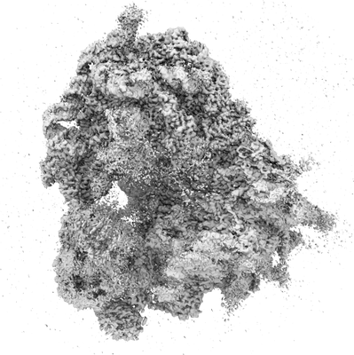 emd_13112.png emd_13112.png | 87.5 KB | ||
| Filedesc metadata | emd-13112.cif.gz | 19.3 KB | ||
| Archive directory |  http://ftp.pdbj.org/pub/emdb/structures/EMD-13112ftp://ftp.pdbj.org/pub/emdb/structures/EMD-13112 http://ftp.pdbj.org/pub/emdb/structures/EMD-13112ftp://ftp.pdbj.org/pub/emdb/structures/EMD-13112 | HTTPS FTP |
-Related structure data
| Related structure data |  7oybMC  7oyaC  7oycC  7oydC M: atomic model generated by this map C: citing same article ( |
|---|---|
| Similar structure data |


-Links
| EMDB pages | EMDB (EBI/PDBe) / EMDataResource |
|---|---|
| Related items in Molecule of the Month |


-Map
| File | Download / File: emd_13112.map.gz / Format: CCP4 / Size: 421.9 MB / Type: IMAGE STORED AS FLOATING POINT NUMBER (4 BYTES) | ||||||||||||||||||||||||||||||||||||
|---|---|---|---|---|---|---|---|---|---|---|---|---|---|---|---|---|---|---|---|---|---|---|---|---|---|---|---|---|---|---|---|---|---|---|---|---|---|
| Projections & slices | Image control
Images are generated by Spider. | ||||||||||||||||||||||||||||||||||||
| Voxel size | X=Y=Z: 1.06 Å | ||||||||||||||||||||||||||||||||||||
| Density |
| ||||||||||||||||||||||||||||||||||||
| Symmetry | Space group: 1 | ||||||||||||||||||||||||||||||||||||
| Details | EMDB XML:
|
 Z (Sec.)
 Y (Row.)
 X (Col.)


-Supplemental data
- Sample components
+Entire : 80S ribosome from 6 hpf zebrafish embryos
+Supramolecule #1: 80S ribosome from 6 hpf zebrafish embryos
+Macromolecule #1: 18S rRNA
+Macromolecule #2: 28S rRNA
+Macromolecule #3: 5S rRNA
+Macromolecule #4: 5.8S rRNA
+Macromolecule #5: 60S ribosomal protein L8
+Macromolecule #6: 40S ribosomal protein SA
+Macromolecule #7: Ribosomal protein L3
+Macromolecule #8: 40S ribosomal protein S3a
+Macromolecule #9: Ribosomal protein L4
+Macromolecule #10: 40S ribosomal protein S2
+Macromolecule #11: Ribosomal protein L5b
+Macromolecule #12: DNA-(apurinic or apyrimidinic site) lyase
+Macromolecule #13: 60S ribosomal protein L6
+Macromolecule #14: 40S ribosomal protein S4, X isoform
+Macromolecule #15: Ribosomal protein L7
+Macromolecule #16: Ribosomal protein S5
+Macromolecule #17: 60S ribosomal protein L7a
+Macromolecule #18: 40S ribosomal protein S6
+Macromolecule #19: 60S ribosomal protein L9
+Macromolecule #20: 40S ribosomal protein S7
+Macromolecule #21: 60S ribosomal protein L10
+Macromolecule #22: 40S ribosomal protein S8
+Macromolecule #23: 60S ribosomal protein L11
+Macromolecule #24: 40S ribosomal protein S9
+Macromolecule #25: Ribosomal protein S10
+Macromolecule #26: 60S ribosomal protein L13
+Macromolecule #27: 40S ribosomal protein S11
+Macromolecule #28: 60S ribosomal protein L14
+Macromolecule #29: Ribosomal protein L15
+Macromolecule #30: 40S ribosomal protein S13
+Macromolecule #31: 60S ribosomal protein L13a
+Macromolecule #32: Ribosomal protein S14
+Macromolecule #33: 60S ribosomal protein L17
+Macromolecule #34: 40S ribosomal protein S15
+Macromolecule #35: Ribosomal protein L18
+Macromolecule #36: Ribosomal protein S16
+Macromolecule #37: 60S ribosomal protein L19
+Macromolecule #38: 40S ribosomal protein S17
+Macromolecule #39: 60S ribosomal protein L18a
+Macromolecule #40: 40S ribosomal protein S18
+Macromolecule #41: 60S ribosomal protein L21
+Macromolecule #42: 40S ribosomal protein S19
+Macromolecule #43: Ribosomal protein L22
+Macromolecule #44: 40S ribosomal protein S20
+Macromolecule #45: 60S ribosomal protein L23
+Macromolecule #46: 40S ribosomal protein S21
+Macromolecule #47: 60S ribosomal protein L24
+Macromolecule #48: 40S ribosomal protein S15a
+Macromolecule #49: Ribosomal protein L23a
+Macromolecule #50: 40S ribosomal protein S23
+Macromolecule #51: ATPase H+ transporting V0 subunit e1
+Macromolecule #52: 40S ribosomal protein S24
+Macromolecule #53: 60S ribosomal protein L27
+Macromolecule #54: 40S ribosomal protein S25
+Macromolecule #55: 60S ribosomal protein L27a
+Macromolecule #56: 40S ribosomal protein S26
+Macromolecule #57: 60S ribosomal protein L29
+Macromolecule #58: 40S ribosomal protein S27
+Macromolecule #59: 60S ribosomal protein L30
+Macromolecule #60: 40S ribosomal protein S28
+Macromolecule #61: 60S ribosomal protein L31
+Macromolecule #62: 40S ribosomal protein S29
+Macromolecule #63: Ribosomal protein L32
+Macromolecule #64: 40S ribosomal protein S30
+Macromolecule #65: 60S ribosomal protein L35a
+Macromolecule #66: 60S ribosomal protein L34
+Macromolecule #67: Guanine nucleotide-binding protein subunit beta-2-like 1
+Macromolecule #68: 60S ribosomal protein L35
+Macromolecule #69: 60S ribosomal protein L36
+Macromolecule #70: Ribosomal protein L37
+Macromolecule #71: 60S ribosomal protein L38
+Macromolecule #72: Ribosomal protein L39
+Macromolecule #73: 60S ribosomal protein L40
+Macromolecule #74: Rpl41
+Macromolecule #75: 60S ribosomal protein L36a
+Macromolecule #76: Zgc:171772
+Macromolecule #77: 60S ribosomal protein L28
+Macromolecule #78: MAGNESIUM ION
+Macromolecule #79: ZINC ION
-Experimental details
-Structure determination
| Method | cryo EM |
|---|---|
 Processing Processing | single particle reconstruction |
| Aggregation state | particle |
-Sample preparation
| Buffer | pH: 7.6 Component:
| ||||||||||||||||||
|---|---|---|---|---|---|---|---|---|---|---|---|---|---|---|---|---|---|---|---|
| Grid | Model: Quantifoil R3.5/1 / Material: COPPER / Mesh: 200 / Support film - Material: CARBON / Support film - topology: CONTINUOUS / Support film - Film thickness: 2 / Pretreatment - Type: GLOW DISCHARGE / Pretreatment - Time: 60 sec. | ||||||||||||||||||
| Vitrification | Cryogen name: ETHANE / Chamber humidity: 70 % / Instrument: LEICA EM GP |
- Electron microscopy
| Microscope | FEI TITAN KRIOS |
|---|---|
| Image recording | Film or detector model: FEI FALCON III (4k x 4k) / Number grids imaged: 1 / Number real images: 11860 / Average electron dose: 48.3 e/Å2 |
| Electron beam | Acceleration voltage: 300 kV / Electron source:  FIELD EMISSION GUN FIELD EMISSION GUN |
| Electron optics | Illumination mode: FLOOD BEAM / Imaging mode: BRIGHT FIELD / Cs: 2.7 mm |
| Sample stage | Specimen holder model: FEI TITAN KRIOS AUTOGRID HOLDER / Cooling holder cryogen: NITROGEN |
| Experimental equipment |  Model: Titan Krios / Image courtesy: FEI Company |
+Image processing
-Atomic model buiding 1
| Refinement | Protocol: OTHER |
|---|---|
| Output model | PDB-7oyb: |
