ムービー
ムービー コントローラー


+ データを開く

- 基本情報
基本情報
| 登録情報 | データベース: EMDB / ID: EMD-12901 | |||||||||
|---|---|---|---|---|---|---|---|---|---|---|
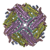
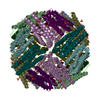
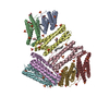
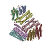
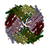
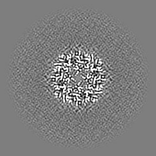
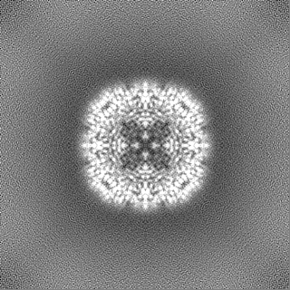
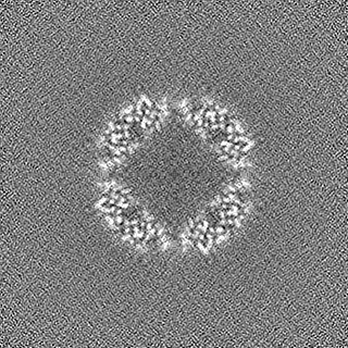
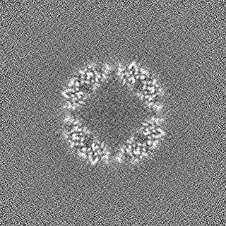
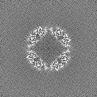
| タイトル | Cryo-EM structure of pyrococcus furiosus apoferritin in nanofluidic channels | |||||||||
 マップデータ マップデータ | ||||||||||
 試料 試料 |
| |||||||||
 キーワード キーワード | Iron Storage / METAL TRANSPORT | |||||||||
| 機能・相同性 |  機能・相同性情報 機能・相同性情報ferroxidase activity / ferric iron binding / iron ion transport / ferrous iron binding / intracellular iron ion homeostasis / identical protein binding / cytosol 類似検索 - 分子機能 | |||||||||
| 生物種 |   Pyrococcus furiosus COM1 (古細菌) Pyrococcus furiosus COM1 (古細菌) | |||||||||
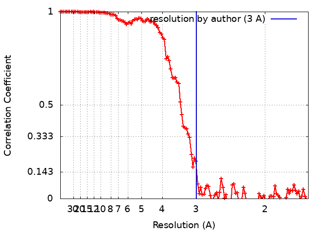
| 手法 | 単粒子再構成法 / クライオ電子顕微鏡法 / 解像度: 3.0 Å | |||||||||
 データ登録者 データ登録者 | Huber ST / Sarajlic E / Huijink R / Evers WH / Jakobi AJ | |||||||||
| 資金援助 | European Union,  オランダ, 2件 オランダ, 2件
| |||||||||
 引用 引用 | ジャーナル: Elife / 年: 2022 タイトル: Nanofluidic chips for cryo-EM structure determination from picoliter sample volumes. 著者: Stefan T Huber / Edin Sarajlic / Roeland Huijink / Felix Weis / Wiel H Evers / Arjen J Jakobi /  要旨: Cryogenic electron microscopy has become an essential tool for structure determination of biological macromolecules. In practice, the difficulty to reliably prepare samples with uniform ice thickness ...Cryogenic electron microscopy has become an essential tool for structure determination of biological macromolecules. In practice, the difficulty to reliably prepare samples with uniform ice thickness still represents a barrier for routine high-resolution imaging and limits the current throughput of the technique. We show that a nanofluidic sample support with well-defined geometry can be used to prepare cryo-EM specimens with reproducible ice thickness from picoliter sample volumes. The sample solution is contained in electron-transparent nanochannels that provide uniform thickness gradients without further optimisation and eliminate the potentially destructive air-water interface. We demonstrate the possibility to perform high-resolution structure determination with three standard protein specimens. Nanofabricated sample supports bear potential to automate the cryo-EM workflow, and to explore new frontiers for cryo-EM applications such as time-resolved imaging and high-throughput screening. #1: ジャーナル: Biorxiv / 年: 2021タイトル: Nanofluidic chips for cryo-EM structure determination from picoliter sample volumes 著者: Huber ST / Sarajlic E / Huijink R / Weis F / Evers WH / Jakobi AJ | |||||||||
| 履歴 |
|
- 構造の表示
構造の表示
| ムービー |
ムービービューア |
|---|---|
| 構造ビューア | EMマップ: SurfViewMolmilJmol/JSmol |
| 添付画像 |

- ダウンロードとリンク
-EMDBアーカイブ
| マップデータ | emd_12901.map.gz | 118 MB | EMDBマップデータ形式 | |
|---|---|---|---|---|
| ヘッダ (付随情報) | emd-12901-v30.xmlemd-12901.xml | 21.4 KB 21.4 KB | 表示 表示 | EMDBヘッダ |
| FSC (解像度算出) | emd_12901_fsc.xml | 11.1 KB | 表示 | FSCデータファイル |
| 画像 | 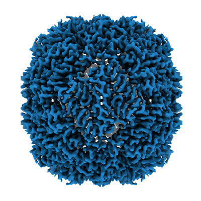 emd_12901.png emd_12901.png | 197.8 KB | ||
| マスクデータ | emd_12901_msk_1.map | 125 MB | マスクマップ | |
| Filedesc metadata | emd-12901.cif.gz | 5.9 KB | ||
| その他 | emd_12901_additional_1.map.gzemd_12901_additional_2.map.gzemd_12901_half_map_1.map.gzemd_12901_half_map_2.map.gz | 62.4 MB 9.9 MB 116 MB 116 MB | ||
| アーカイブディレクトリ |  http://ftp.pdbj.org/pub/emdb/structures/EMD-12901ftp://ftp.pdbj.org/pub/emdb/structures/EMD-12901 http://ftp.pdbj.org/pub/emdb/structures/EMD-12901ftp://ftp.pdbj.org/pub/emdb/structures/EMD-12901 | HTTPS FTP |
-関連構造データ
| 関連構造データ |  7ohfMC M: このマップから作成された原子モデル C: 同じ文献を引用 ( |
|---|---|
| 類似構造データ | |
| 電子顕微鏡画像生データ | EMPIAR-10708 (タイトル: Apoferritin, TMV and T20S proteasome in nanofluidic channels Data size: 308.3 Data #1: Unaligned multi-frame movies of pyrococcus furiosus apoferritin in silicon nitride nanochannels [micrographs - multiframe] Data #2: Multi-frame movies of T20S proteasome in silicon nitride nanochannels [micrographs - multiframe] Data #3: Multi-frame movies of TMV in silicon nitride nanochannels [micrographs - multiframe]) |


-リンク
| EMDBのページ | EMDB (EBI/PDBe) / EMDataResource |
|---|---|
| 「今月の分子」の関連する項目 |


-マップ
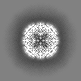
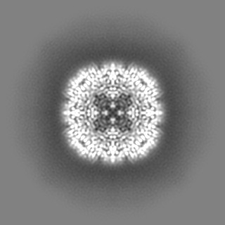
| ファイル | ダウンロード / ファイル: emd_12901.map.gz / 形式: CCP4 / 大きさ: 125 MB / タイプ: IMAGE STORED AS FLOATING POINT NUMBER (4 BYTES) | ||||||||||||||||||||||||||||||||||||||||||||||||||||||||||||
|---|---|---|---|---|---|---|---|---|---|---|---|---|---|---|---|---|---|---|---|---|---|---|---|---|---|---|---|---|---|---|---|---|---|---|---|---|---|---|---|---|---|---|---|---|---|---|---|---|---|---|---|---|---|---|---|---|---|---|---|---|---|
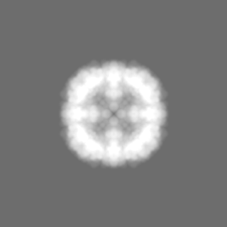
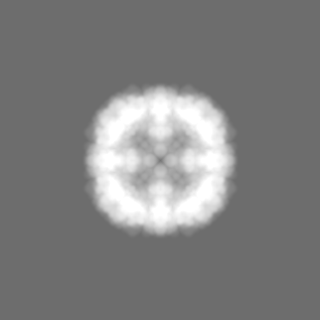
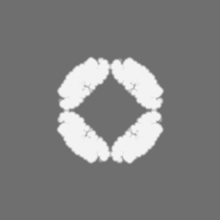
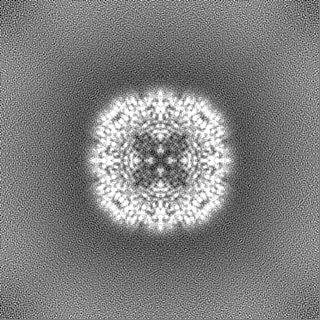
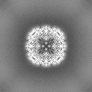
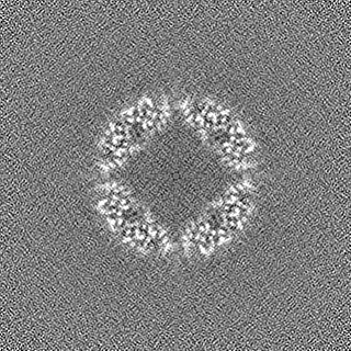
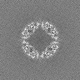
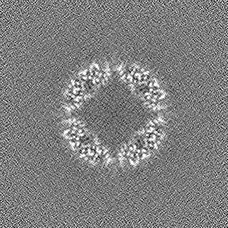
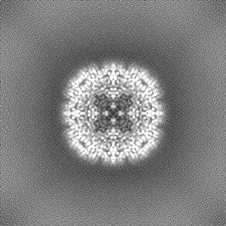
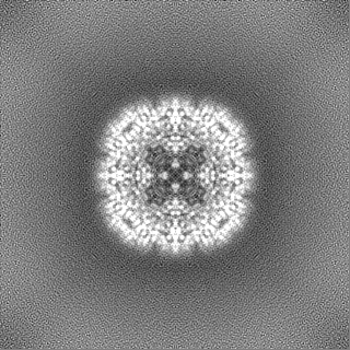
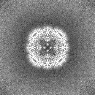
| 投影像・断面図 | 画像のコントロール
画像は Spider により作成 | ||||||||||||||||||||||||||||||||||||||||||||||||||||||||||||
| ボクセルのサイズ | X=Y=Z: 0.8127 Å | ||||||||||||||||||||||||||||||||||||||||||||||||||||||||||||
| 密度 |
| ||||||||||||||||||||||||||||||||||||||||||||||||||||||||||||
| 対称性 | 空間群: 1 | ||||||||||||||||||||||||||||||||||||||||||||||||||||||||||||
| 詳細 | EMDB XML:
CCP4マップ ヘッダ情報:
| ||||||||||||||||||||||||||||||||||||||||||||||||||||||||||||
 Z (Sec.)
 Y (Row.)
 X (Col.)


-添付データ
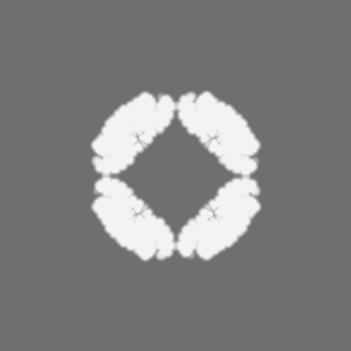
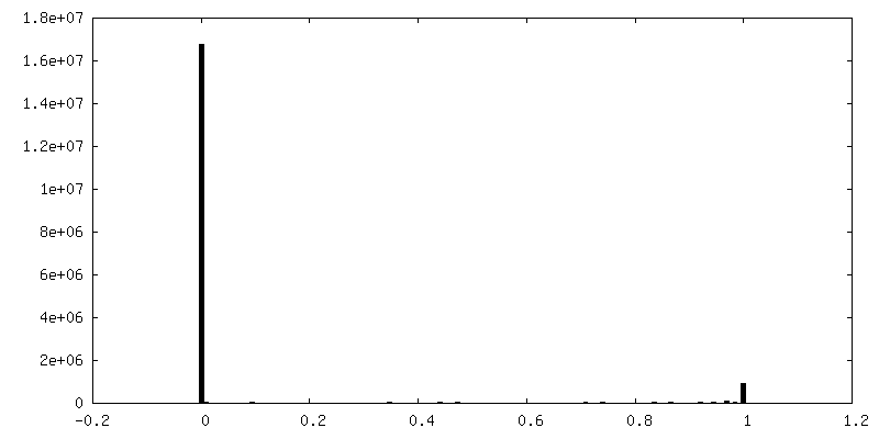
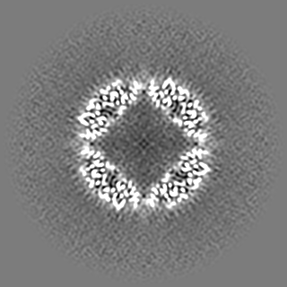
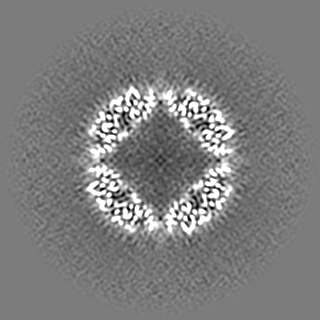
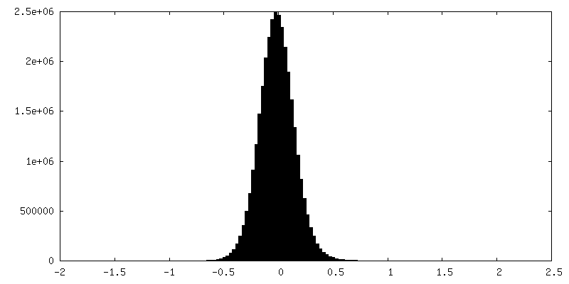
-マスク #1
| ファイル | emd_12901_msk_1.map | ||||||||||||
|---|---|---|---|---|---|---|---|---|---|---|---|---|---|
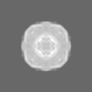
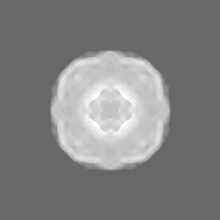
| 投影像・断面図 |
| ||||||||||||
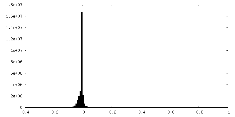
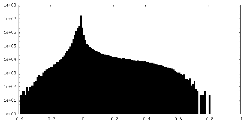
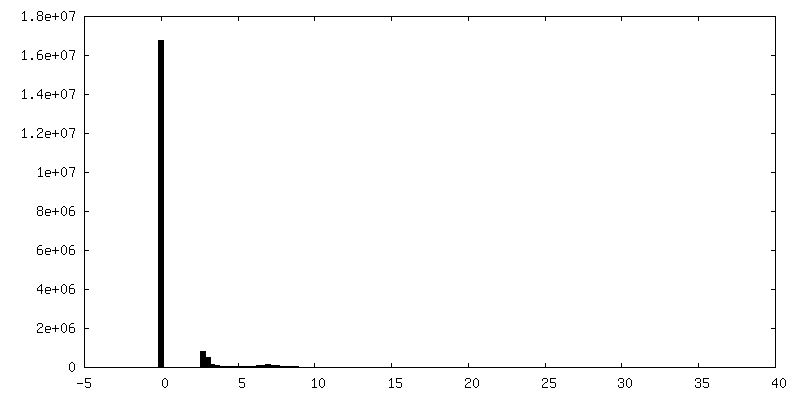
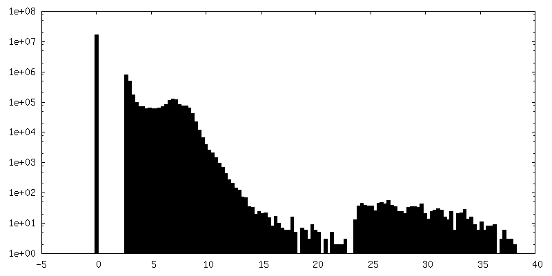
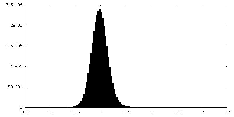
| 密度ヒストグラム |


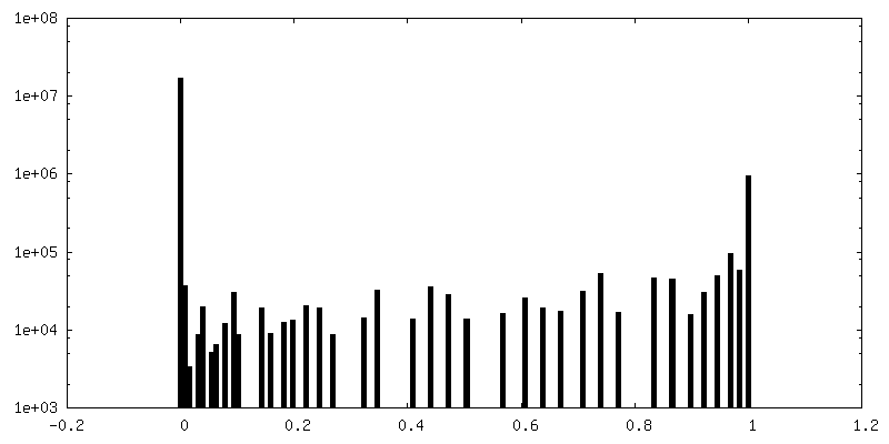
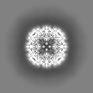
-追加マップ: #1
| ファイル | emd_12901_additional_1.map | ||||||||||||
|---|---|---|---|---|---|---|---|---|---|---|---|---|---|
| 投影像・断面図 |
| ||||||||||||
| 密度ヒストグラム |

-追加マップ: #2
| ファイル | emd_12901_additional_2.map | ||||||||||||
|---|---|---|---|---|---|---|---|---|---|---|---|---|---|
| 投影像・断面図 |
| ||||||||||||
| 密度ヒストグラム |
-ハーフマップ: #1
| ファイル | emd_12901_half_map_1.map | ||||||||||||
|---|---|---|---|---|---|---|---|---|---|---|---|---|---|
| 投影像・断面図 |
| ||||||||||||
| 密度ヒストグラム |

-ハーフマップ: #2
| ファイル | emd_12901_half_map_2.map | ||||||||||||
|---|---|---|---|---|---|---|---|---|---|---|---|---|---|
| 投影像・断面図 |
| ||||||||||||
| 密度ヒストグラム |
- 試料の構成要素
-全体 : 24-mer of pyrococcus furiosus apoferritin
| 全体 | 名称: 24-mer of pyrococcus furiosus apoferritin |
|---|---|
| 要素 |
|
-超分子 #1: 24-mer of pyrococcus furiosus apoferritin
| 超分子 | 名称: 24-mer of pyrococcus furiosus apoferritin / タイプ: complex / ID: 1 / 親要素: 0 / 含まれる分子: all |
|---|---|
| 由来(天然) | 生物種: Pyrococcus furiosus COM1 (古細菌) |
| 分子量 | 理論値: 492 KDa |
-分子 #1: Ferritin
| 分子 | 名称: Ferritin / タイプ: protein_or_peptide / ID: 1 / コピー数: 1 / 光学異性体: LEVO |
|---|---|
| 由来(天然) | 生物種: Pyrococcus furiosus COM1 (古細菌) |
| 分子量 | 理論値: 20.535395 KDa |
| 組換発現 | 生物種:  |
| 配列 | 文字列: MAMLSERMLK ALNDQLNREL YSAYLYFAMA AYFEDLGLEG FANWMKAQAE EEIGHALRFY NYIYDRNGRV ELDEIPKPPK EWESPLKAF EAAYEHEKFI SKSIYELAAL AEEEKDYSTR AFLEWFINEQ VEEEASVKKI LDKLKFAKDS PQILFMLDKE L SARAPKLP GLLMQGGE UniProtKB: Ferritin |
-実験情報
-構造解析
| 手法 | クライオ電子顕微鏡法 |
|---|---|
 解析 解析 | 単粒子再構成法 |
| 試料の集合状態 | particle |
-試料調製
| 濃度 | 3.4 mg/mL | |||||||||
|---|---|---|---|---|---|---|---|---|---|---|
| 緩衝液 | pH: 7.5 構成要素:
| |||||||||
| グリッド | モデル: Homemade / 材質: SILICON NITRIDE | |||||||||
| 凍結 | 凍結剤: ETHANE / 装置: LEICA PLUNGER 詳細: The sample was filled into cryoChips through the cantilever and then transferred within ~10 seconds to the Leica plunger for freezing.. | |||||||||
| 詳細 | The sample was filled into nanofluidic channels. |
- 電子顕微鏡法
| 顕微鏡 | FEI TITAN KRIOS |
|---|---|
| 撮影 | フィルム・検出器のモデル: GATAN K2 SUMMIT (4k x 4k) 検出モード: COUNTING / 撮影したグリッド数: 3 / 実像数: 948 / 平均露光時間: 9.0 sec. / 平均電子線量: 63.0 e/Å2 |
| 電子線 | 加速電圧: 300 kV / 電子線源:  FIELD EMISSION GUN FIELD EMISSION GUN |
| 電子光学系 | 照射モード: FLOOD BEAM / 撮影モード: BRIGHT FIELD / Cs: 2.7 mm / 最大 デフォーカス(公称値): -2.0 µm / 最小 デフォーカス(公称値): -1.0 µm |
| 試料ステージ | 試料ホルダーモデル: FEI TITAN KRIOS AUTOGRID HOLDER |
| 実験機器 |  モデル: Titan Krios / 画像提供: FEI Company |