 Movie
Movie Controller


+ Open data

- Basic information
| Entry | Database: PDB / ID: 6txn | ||||||
|---|---|---|---|---|---|---|---|
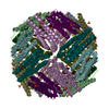
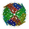
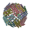
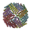
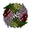
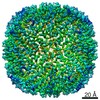
| Title | Crystal structure of thermotoga maritima Ferritin in apo form | ||||||
 Components Components | Ferritin | ||||||
 Keywords Keywords | METAL BINDING PROTEIN / Metal binding / engineered protein | ||||||
| Function / homology |  Function and homology information Function and homology informationbacterial non-heme ferritin / ferroxidase activity / ferric iron binding / iron ion transport / ferrous iron binding / intracellular iron ion homeostasis / identical protein binding / cytoplasm / cytosol Similarity search - Function | ||||||
| Biological species |   Thermotoga maritima (bacteria) Thermotoga maritima (bacteria) | ||||||
| Method |  X-RAY DIFFRACTION / SYNCHROTRON / MOLECULAR REPLACEMENT / Resolution: 2.01 Å X-RAY DIFFRACTION / SYNCHROTRON / MOLECULAR REPLACEMENT / Resolution: 2.01 Å | ||||||
 Authors Authors | Wilk, P. / Grudnik, P. / Kumar, M. / Heddle, J. / Chakraborti, S. | ||||||
| Funding support |  Poland, 1items Poland, 1items
| ||||||
 Citation Citation | Journal: Nanoscale / Year: 2021 Title: A single residue can modulate nanocage assembly in salt dependent ferritin. Authors: Kumar, M. / Markiewicz-Mizera, J. / Janna Olmos, J.D. / Wilk, P. / Grudnik, P. / Biela, A.P. / Jemiola-Rzeminska, M. / Gorecki, A. / Chakraborti, S. / Heddle, J.G. | ||||||
| History |
|
- Structure visualization
| Structure viewer | Molecule: MolmilJmol/JSmol |
|---|
- Downloads & links
-Download
| PDBx/mmCIF format | 6txn.cif.gz | 525.7 KB | Display | PDBx/mmCIF format |
|---|---|---|---|---|
| PDB format | pdb6txn.ent.gz | 444.5 KB | Display | PDB format |
| PDBx/mmJSON format | 6txn.json.gz | Tree view | PDBx/mmJSON format | |
| Others |  Other downloads Other downloads |
-Validation report
| Arichive directory | https://data.pdbj.org/pub/pdb/validation_reports/tx/6txnftp://data.pdbj.org/pub/pdb/validation_reports/tx/6txn | HTTPS FTP |
|---|
-Related structure data
| Related structure data |  6txhC  6txiC  6txjC  6txkC  6txlC  6txmC  1vlgS S: Starting model for refinement C: citing same article ( |
|---|---|
| Similar structure data |


-Links
 PDBj
PDBj


- Assembly
| Deposited unit | 
| ||||||||
|---|---|---|---|---|---|---|---|---|---|
| 1 | 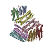
| ||||||||
| Unit cell |
|
-Components
-Protein , 1 types, 8 molecules ABCDEFGH
| #1: Protein | Mass: 19401.840 Da / Num. of mol.: 8 Source method: isolated from a genetically manipulated source Source: (gene. exp.) Thermotoga maritima (strain ATCC 43589 / MSB8 / DSM 3109 / JCM 10099) (bacteria)Gene: TM_1128 / Production host: |
|---|
-Non-polymers , 5 types, 677 molecules 


| #2: Chemical | ChemComp-GOL /  Mass: 92.094 Da / Num. of mol.: 10 / Source method: obtained synthetically / Formula: C3H8O3 / Feature type: SUBJECT OF INVESTIGATION Mass: 92.094 Da / Num. of mol.: 10 / Source method: obtained synthetically / Formula: C3H8O3 / Feature type: SUBJECT OF INVESTIGATION#3: Chemical | ChemComp-SO4 /  Mass: 96.063 Da / Num. of mol.: 12 / Source method: obtained synthetically / Formula: SO4 / Feature type: SUBJECT OF INVESTIGATION Mass: 96.063 Da / Num. of mol.: 12 / Source method: obtained synthetically / Formula: SO4 / Feature type: SUBJECT OF INVESTIGATION#4: Chemical | ChemComp-MG /  Mass: 24.305 Da / Num. of mol.: 8 / Source method: obtained synthetically / Formula: Mg / Feature type: SUBJECT OF INVESTIGATION Mass: 24.305 Da / Num. of mol.: 8 / Source method: obtained synthetically / Formula: Mg / Feature type: SUBJECT OF INVESTIGATION#5: Chemical | ChemComp-LFA /  Mass: 282.547 Da / Num. of mol.: 4 / Source method: obtained synthetically / Formula: C20H42 / Feature type: SUBJECT OF INVESTIGATION Mass: 282.547 Da / Num. of mol.: 4 / Source method: obtained synthetically / Formula: C20H42 / Feature type: SUBJECT OF INVESTIGATION#6: Water | ChemComp-HOH / | Mass: 18.015 Da / Num. of mol.: 643 / Source method: isolated from a natural source / Formula: H2O |
|---|
-Details
| Has ligand of interest | Y |
|---|
-Experimental details
-Experiment
| Experiment | Method: X-RAY DIFFRACTION / Number of used crystals: 1 |
|---|
- Sample preparation
| Crystal | Density Matthews: 3.42 Å3/Da / Density % sol: 64.05 % |
|---|---|
| Crystal grow | Temperature: 293 K / Method: vapor diffusion, sitting drop Details: 2 M MgCl2, 2.4 M (NH4)2 SO4, 0.1M MES. pH 6.0. 1ul of 10mg/mL protein was mixed with same amount of well solution, crystals normally appears in 2 days |
-Data collection
| Diffraction | Mean temperature: 100 K / Serial crystal experiment: N |
|---|---|
| Diffraction source | Source: SYNCHROTRON / Site: ESRF  / Beamline: ID23-1 / Wavelength: 0.978631 Å / Beamline: ID23-1 / Wavelength: 0.978631 Å |
| Detector | Type: DECTRIS PILATUS3 S 6M / Detector: PIXEL / Date: Sep 30, 2018 |
| Radiation | Monochromator: 0.978631 / Protocol: SINGLE WAVELENGTH / Monochromatic (M) / Laue (L): M / Scattering type: x-ray |
| Radiation wavelength | Wavelength: 0.978631 Å / Relative weight: 1 |
| Reflection | Resolution: 2.01→49.22 Å / Num. obs: 140330 / % possible obs: 99.9 % / Redundancy: 6.5 % / CC1/2: 0.996 / Rmerge(I) obs: 0.084 / Net I/σ(I): 11 |
| Reflection shell | Resolution: 2.01→7.78 Å / Rmerge(I) obs: 1.526 / Num. unique obs: 13704 / CC1/2: 0.395 / % possible all: 100 |
- Processing
| Software |
| ||||||||||||||||||||||||||||||||||||||||||||||||||||||||||||||||||||||||||||||||||||||||||
|---|---|---|---|---|---|---|---|---|---|---|---|---|---|---|---|---|---|---|---|---|---|---|---|---|---|---|---|---|---|---|---|---|---|---|---|---|---|---|---|---|---|---|---|---|---|---|---|---|---|---|---|---|---|---|---|---|---|---|---|---|---|---|---|---|---|---|---|---|---|---|---|---|---|---|---|---|---|---|---|---|---|---|---|---|---|---|---|---|---|---|---|
| Refinement | Method to determine structure: MOLECULAR REPLACEMENT Starting model: 1vlg Resolution: 2.01→48.276 Å / SU ML: 0.27 / Cross valid method: THROUGHOUT / σ(F): 1.33 / Phase error: 23.83
| ||||||||||||||||||||||||||||||||||||||||||||||||||||||||||||||||||||||||||||||||||||||||||
| Solvent computation | Shrinkage radii: 0.9 Å / VDW probe radii: 1.11 Å | ||||||||||||||||||||||||||||||||||||||||||||||||||||||||||||||||||||||||||||||||||||||||||
| Displacement parameters | Biso max: 343.19 Å2 / Biso mean: 57.4759 Å2 / Biso min: 27.95 Å2 | ||||||||||||||||||||||||||||||||||||||||||||||||||||||||||||||||||||||||||||||||||||||||||
| Refinement step | Cycle: final / Resolution: 2.01→48.276 Å
| ||||||||||||||||||||||||||||||||||||||||||||||||||||||||||||||||||||||||||||||||||||||||||
| LS refinement shell | Refine-ID: X-RAY DIFFRACTION / Rfactor Rfree error: 0
|
