ムービー
ムービー コントローラー


+ データを開く

- 基本情報
基本情報
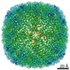
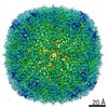
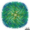
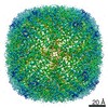
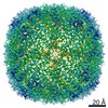
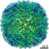
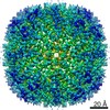
| 登録情報 | データベース: EMDB / ID: EMD-12358 | |||||||||
|---|---|---|---|---|---|---|---|---|---|---|
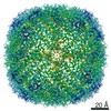
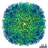
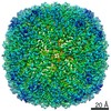
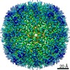
| タイトル | Mouse heavy chain apoferritin collected on cryoARM300 with coma-corrected beam-image shift | |||||||||
 マップデータ マップデータ | ||||||||||
 試料 試料 |
| |||||||||
| 機能・相同性 |  機能・相同性情報 機能・相同性情報Iron uptake and transport / Golgi Associated Vesicle Biogenesis / negative regulation of ferroptosis / iron ion sequestering activity / autolysosome / ferroxidase / intracellular sequestering of iron ion / ferroxidase activity / negative regulation of fibroblast proliferation / endocytic vesicle lumen ...Iron uptake and transport / Golgi Associated Vesicle Biogenesis / negative regulation of ferroptosis / iron ion sequestering activity / autolysosome / ferroxidase / intracellular sequestering of iron ion / ferroxidase activity / negative regulation of fibroblast proliferation / endocytic vesicle lumen / Neutrophil degranulation / ferric iron binding / ferrous iron binding / iron ion transport / iron ion binding / immune response / negative regulation of cell population proliferation / mitochondrion / extracellular region / identical protein binding / membrane / cytoplasm / cytosol 類似検索 - 分子機能 | |||||||||
| 生物種 |  | |||||||||
| 手法 | 単粒子再構成法 / クライオ電子顕微鏡法 / 解像度: 1.7 Å | |||||||||
 データ登録者 データ登録者 | Efremov R / Stroobants A | |||||||||
| 資金援助 |  ベルギー, 1件 ベルギー, 1件
| |||||||||
 引用 引用 | ジャーナル: Acta Crystallogr D Struct Biol / 年: 2021 タイトル: Coma-corrected rapid single-particle cryo-EM data collection on the CRYO ARM 300. 著者: Rouslan G Efremov / Annelore Stroobants / 要旨: Single-particle cryogenic electron microscopy has recently become a major method for determining the structures of proteins and protein complexes. This has markedly increased the demand for ...Single-particle cryogenic electron microscopy has recently become a major method for determining the structures of proteins and protein complexes. This has markedly increased the demand for throughput of high-resolution electron microscopes, which are required to produce high-resolution images at high rates. An increase in data-collection throughput can be achieved by using large beam-image shifts combined with off-axis coma correction, enabling the acquisition of multiple images from a large area of the EM grid without moving the microscope stage. Here, the optical properties of the JEOL CRYO ARM 300 electron microscope equipped with a K3 camera were characterized under off-axis illumination conditions. It is shown that efficient coma correction can be achieved for beam-image shifts with an amplitude of at least 10 µm, enabling a routine throughput for data collection of between 6000 and 9000 images per day. Use of the benchmark for the rapid data-collection procedure (with beam-image shifts of up to 7 µm) on apoferritin resulted in a reconstruction at a resolution of 1.7 Å. This demonstrates that the rapid automated acquisition of high-resolution micrographs is possible using a CRYO ARM 300. | |||||||||
| 履歴 |
|
- 構造の表示
構造の表示
| ムービー |
ムービービューア |
|---|---|
| 構造ビューア | EMマップ: SurfViewMolmilJmol/JSmol |
| 添付画像 |

- ダウンロードとリンク
-EMDBアーカイブ
| マップデータ | emd_12358.map.gz | 15.4 MB | EMDBマップデータ形式 | |
|---|---|---|---|---|
| ヘッダ (付随情報) | emd-12358-v30.xmlemd-12358.xml | 16.8 KB 16.8 KB | 表示 表示 | EMDBヘッダ |
| FSC (解像度算出) | emd_12358_fsc.xml | 9.9 KB | 表示 | FSCデータファイル |
| 画像 | 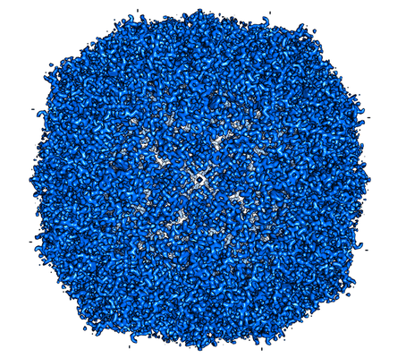 emd_12358.png emd_12358.png | 314.6 KB | ||
| マスクデータ | emd_12358_msk_1.map | 83.7 MB | マスクマップ | |
| その他 | emd_12358_half_map_1.map.gzemd_12358_half_map_2.map.gz | 61.1 MB 61.1 MB | ||
| アーカイブディレクトリ |  http://ftp.pdbj.org/pub/emdb/structures/EMD-12358ftp://ftp.pdbj.org/pub/emdb/structures/EMD-12358 http://ftp.pdbj.org/pub/emdb/structures/EMD-12358ftp://ftp.pdbj.org/pub/emdb/structures/EMD-12358 | HTTPS FTP |
-検証レポート
| 文書・要旨 | emd_12358_validation.pdf.gz | 376.8 KB | 表示 | EMDB検証レポート |
|---|---|---|---|---|
| 文書・詳細版 | emd_12358_full_validation.pdf.gz | 376 KB | 表示 | |
| XML形式データ | emd_12358_validation.xml.gz | 15.9 KB | 表示 | |
| アーカイブディレクトリ | https://ftp.pdbj.org/pub/emdb/validation_reports/EMD-12358ftp://ftp.pdbj.org/pub/emdb/validation_reports/EMD-12358 | HTTPS FTP |
-関連構造データ
| 類似構造データ | |
|---|---|
| 電子顕微鏡画像生データ | EMPIAR-10639 (タイトル: Single particle cryo-EM dataset of mouse heavy chain apoferritin collected on cryoARM300 with beam-image shift of 7 um Data size: 695.6 Data #1: Unaligned multi frame micrographs of mouse heavy chain apoferritin collected on cryoARM300 with image shift 7um [micrographs - multiframe]) |


-リンク
| EMDBのページ | EMDB (EBI/PDBe) / EMDataResource |
|---|---|
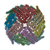
| 「今月の分子」の関連する項目 |


-マップ
| ファイル | ダウンロード / ファイル: emd_12358.map.gz / 形式: CCP4 / 大きさ: 83.7 MB / タイプ: IMAGE STORED AS FLOATING POINT NUMBER (4 BYTES) | ||||||||||||||||||||||||||||||||||||||||||||||||||||||||||||
|---|---|---|---|---|---|---|---|---|---|---|---|---|---|---|---|---|---|---|---|---|---|---|---|---|---|---|---|---|---|---|---|---|---|---|---|---|---|---|---|---|---|---|---|---|---|---|---|---|---|---|---|---|---|---|---|---|---|---|---|---|---|
| ボクセルのサイズ | X=Y=Z: 0.753 Å | ||||||||||||||||||||||||||||||||||||||||||||||||||||||||||||
| 密度 |
| ||||||||||||||||||||||||||||||||||||||||||||||||||||||||||||
| 対称性 | 空間群: 1 | ||||||||||||||||||||||||||||||||||||||||||||||||||||||||||||
| 詳細 | EMDB XML:
CCP4マップ ヘッダ情報:
| ||||||||||||||||||||||||||||||||||||||||||||||||||||||||||||
-添付データ
-マスク #1
| ファイル | emd_12358_msk_1.map | ||||||||||||
|---|---|---|---|---|---|---|---|---|---|---|---|---|---|
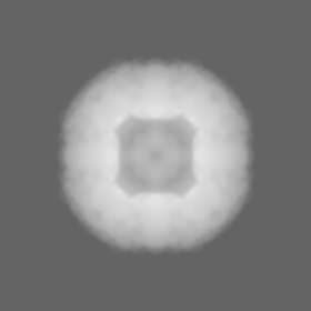
| 投影像・断面図 |
| ||||||||||||
| 密度ヒストグラム |
 Z
Z Y
Y X
X


-ハーフマップ: #1
| ファイル | emd_12358_half_map_1.map | ||||||||||||
|---|---|---|---|---|---|---|---|---|---|---|---|---|---|
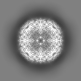
| 投影像・断面図 |
| ||||||||||||
| 密度ヒストグラム |


-ハーフマップ: #2
| ファイル | emd_12358_half_map_2.map | ||||||||||||
|---|---|---|---|---|---|---|---|---|---|---|---|---|---|
| 投影像・断面図 |
| ||||||||||||
| 密度ヒストグラム |


- 試料の構成要素
-全体 : mouse heavy chain apoferritin
| 全体 | 名称: mouse heavy chain apoferritin |
|---|---|
| 要素 |
|
-超分子 #1: mouse heavy chain apoferritin
| 超分子 | 名称: mouse heavy chain apoferritin / タイプ: complex / ID: 1 / 親要素: 0 / 含まれる分子: all / 詳細: Wilde type, octamer |
|---|---|
| 由来(天然) | 生物種: |
| 組換発現 | 生物種:  |
| 分子量 | 理論値: 506 KDa |
-分子 #1: mouse heavy chain apoferritin
| 分子 | 名称: mouse heavy chain apoferritin / タイプ: protein_or_peptide / ID: 1 / 光学異性体: LEVO / EC番号: ferroxidase |
|---|---|
| 由来(天然) | 生物種: |
| 組換発現 | 生物種: |
| 配列 | 文字列: MTTASPSQVR QNYHQDAEAA INRQINLELY ASYVYLSMSC YFDRDDVALK NFAKYFLHQS HEEREHAEK LMKLQNQRGG RIFLQDIKKP DRDDWESGLN AMECALHLEK SVNQSLLELH K LATDKNDP HLCDFIETYY LSEQVKSIKE LGDHVTNLRK MGAPEAGMAE YLFDKHTLGH GD ES |
-実験情報
-構造解析
| 手法 | クライオ電子顕微鏡法 |
|---|---|
 解析 解析 | 単粒子再構成法 |
| 試料の集合状態 | particle |
-試料調製
| 濃度 | 3.6 mg/mL |
|---|---|
| 緩衝液 | pH: 7.5 / 詳細: 20 mM Hepes pH 7.5, 300 mM NaCl, 1mM TCEP |
| グリッド | モデル: Quantifoil / 材質: COPPER / メッシュ: 200 / 支持フィルム - 材質: CARBON / 支持フィルム - トポロジー: HOLEY / 前処理 - タイプ: GLOW DISCHARGE |
| 凍結 | 凍結剤: ETHANE / チャンバー内湿度: 98 % / チャンバー内温度: 298 K / 装置: GATAN CRYOPLUNGE 3 / 詳細: 5 seconds blotting. |
- 電子顕微鏡法
| 顕微鏡 | JEOL CRYO ARM 300 |
|---|---|
| アライメント法 | Coma free - Residual tilt: 0.9 mrad |
| 特殊光学系 | エネルギーフィルター - 名称: In-column Omega Filter エネルギーフィルター - スリット幅: 20 eV |
| 撮影 | フィルム・検出器のモデル: GATAN K3 (6k x 4k) / 検出モード: COUNTING / 撮影したグリッド数: 1 / 実像数: 2639 / 平均露光時間: 3.37 sec. / 平均電子線量: 59.0 e/Å2 |
| 電子線 | 加速電圧: 300 kV / 電子線源:  FIELD EMISSION GUN FIELD EMISSION GUN |
| 電子光学系 | 照射モード: FLOOD BEAM / 撮影モード: BRIGHT FIELD / Cs: 2.55 mm |
| 試料ステージ | 試料ホルダーモデル: JEOL CRYOSPECPORTER / ホルダー冷却材: NITROGEN |
+画像解析

-原子モデル構築 1
| 初期モデル | PDB ID: |
|---|---|
| 精密化 | 空間: REAL / プロトコル: OTHER / 当てはまり具合の基準: correlation coefficient |