 Movie
Movie Controller

[English] 日本語
 Yorodumi
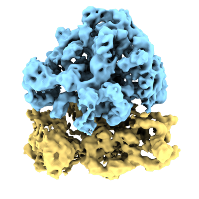
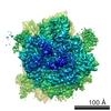
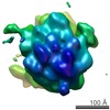
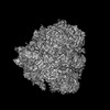
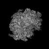
- EMDB-10211: Subtomogram average of E. coli 70S ribosome from FISE tilt series -
+ Open data

- Basic information
| Entry | Database: EMDB / ID: EMD-10211 | |||||||||
|---|---|---|---|---|---|---|---|---|---|---|
| Title | Subtomogram average of E. coli 70S ribosome from FISE tilt series | |||||||||
 Map data Map data | ||||||||||
 Sample Sample |
| |||||||||
| Biological species |  | |||||||||
| Method | subtomogram averaging / cryo EM / Resolution: 9.0 Å | |||||||||
 Authors Authors | Eisenstein F / Danev R / Pilhofer M | |||||||||
 Citation Citation | Journal: J Struct Biol / Year: 2019 Title: Improved applicability and robustness of fast cryo-electron tomography data acquisition. Authors: Fabian Eisenstein / Radostin Danev / Martin Pilhofer /   Abstract: The power of cryo-electron tomography (cryoET) lies in its capability to characterize macromolecules in their cellular context. Structure determination by cryoET, however, is time-consuming compared ...The power of cryo-electron tomography (cryoET) lies in its capability to characterize macromolecules in their cellular context. Structure determination by cryoET, however, is time-consuming compared to single particle approaches. A recent study reported significant acceleration of data acquisition by a fast-incremental single-exposure (FISE) tilt series scheme. Here we improved the method and evaluated its efficiency and performance. We show that (1) FISE combined with the latest generation of direct electron detectors speeds up collection considerably, (2) previous generation (pre-2017) double-tilt axis Titan Krios holders are also suitable for FISE data acquisition, (3) x, y and z-specimen shifts can be compensated for, and (4) FISE tilt series data can generate averages of sub-nanometer resolution. These advances will allow for a widespread adoption of cryoET for high-throughput in situ studies and high-resolution structure determination across different biological research disciplines. #2: Journal: J.Struct.Biol. / Year: 2019Title: Improved applicability and robustness of fast cryo-electron tomography data acquisition Authors: Eisenstein F / Danev R / Pilhofer M | |||||||||
| History |
|
- Structure visualization
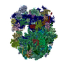
| Movie |
 Movie viewer Movie viewer |
|---|---|
| Structure viewer | EM map: SurfViewMolmilJmol/JSmol |
| Supplemental images |
- Downloads & links
-EMDB archive
| Map data | emd_10211.map.gz | 5.5 MB | EMDB map data format | |
|---|---|---|---|---|
| Header (meta data) | emd-10211-v30.xmlemd-10211.xml | 11.5 KB 11.5 KB | Display Display | EMDB header |
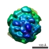
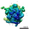
| Images |  emd_10211.png emd_10211.png | 167.8 KB | ||
| Archive directory |  http://ftp.pdbj.org/pub/emdb/structures/EMD-10211ftp://ftp.pdbj.org/pub/emdb/structures/EMD-10211 http://ftp.pdbj.org/pub/emdb/structures/EMD-10211ftp://ftp.pdbj.org/pub/emdb/structures/EMD-10211 | HTTPS FTP |
-Related structure data
| Similar structure data | |
|---|---|
| EM raw data | EMPIAR-10304 (Title: Improved applicability and robustness of fast cryo-electron tomography data acquisition Data size: 21.6 / Data #1: FISEtiltseries [tilt series]) |

-Links
| EMDB pages | EMDB (EBI/PDBe) / EMDataResource |
|---|---|
| Related items in Molecule of the Month |

-Map
| File | Download / File: emd_10211.map.gz / Format: CCP4 / Size: 48.9 MB / Type: IMAGE STORED AS FLOATING POINT NUMBER (4 BYTES) | ||||||||||||||||||||||||||||||||||||||||||||||||||||||||||||
|---|---|---|---|---|---|---|---|---|---|---|---|---|---|---|---|---|---|---|---|---|---|---|---|---|---|---|---|---|---|---|---|---|---|---|---|---|---|---|---|---|---|---|---|---|---|---|---|---|---|---|---|---|---|---|---|---|---|---|---|---|---|
| Projections & slices | Image control
Images are generated by Spider. | ||||||||||||||||||||||||||||||||||||||||||||||||||||||||||||
| Voxel size | X=Y=Z: 2.1 Å | ||||||||||||||||||||||||||||||||||||||||||||||||||||||||||||
| Density |
| ||||||||||||||||||||||||||||||||||||||||||||||||||||||||||||
| Symmetry | Space group: 1 | ||||||||||||||||||||||||||||||||||||||||||||||||||||||||||||
| Details | EMDB XML:
CCP4 map header:
| ||||||||||||||||||||||||||||||||||||||||||||||||||||||||||||
 Z (Sec.)
 Y (Row.)
 X (Col.)


-Supplemental data
- Sample components
-Entire : 70S ribosome
| Entire | Name: 70S ribosome |
|---|---|
| Components |
|
-Supramolecule #1: 70S ribosome
| Supramolecule | Name: 70S ribosome / type: complex / ID: 1 / Parent: 0 |
|---|---|
| Source (natural) | Organism: |
-Experimental details
-Structure determination
| Method | cryo EM |
|---|---|
 Processing Processing | subtomogram averaging |
| Aggregation state | particle |
-Sample preparation
| Buffer | pH: 7.5 |
|---|---|
| Grid | Model: Quantifoil R2/2 / Material: COPPER / Mesh: 200 |
| Vitrification | Cryogen name: ETHANE / Chamber humidity: 100 % |
- Electron microscopy
| Microscope | FEI TITAN KRIOS |
|---|---|
| Details | single-tilt axis holder |
| Image recording | Film or detector model: GATAN K3 BIOQUANTUM (6k x 4k) / Average exposure time: 0.5 sec. / Average electron dose: 150.0 e/Å2 / Details: FISEtomo SerialEM script |
| Electron beam | Acceleration voltage: 300 kV / Electron source:  FIELD EMISSION GUN FIELD EMISSION GUN |
| Electron optics | Illumination mode: FLOOD BEAM / Imaging mode: BRIGHT FIELD / Cs: 2.7 mm |
| Sample stage | Specimen holder model: FEI TITAN KRIOS AUTOGRID HOLDER / Cooling holder cryogen: NITROGEN |
| Experimental equipment |  Model: Titan Krios / Image courtesy: FEI Company |
