 Movie
Movie Controller


+ Open data

- Basic information
| Entry |  | ||||||||||||||||||
|---|---|---|---|---|---|---|---|---|---|---|---|---|---|---|---|---|---|---|---|
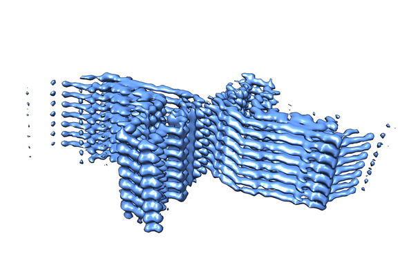
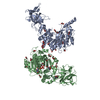
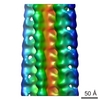
| Title | Two protofilament beta-2-microglobulin amyloid fibril | ||||||||||||||||||
 Map data Map data | Beta-2-microglobulin amyloid fibril - two protofilamets | ||||||||||||||||||
 Sample Sample |
| ||||||||||||||||||
 Keywords Keywords | amyloid / b2m / PROTEIN FIBRIL | ||||||||||||||||||
| Function / homology |  Function and homology information Function and homology informationearly endosome lumen / Nef mediated downregulation of MHC class I complex cell surface expression / DAP12 interactions / Endosomal/Vacuolar pathway / T cell mediated cytotoxicity / Antigen Presentation: Folding, assembly and peptide loading of class I MHC / regulation of iron ion transport / cellular response to iron(III) ion / negative regulation of iron ion transport / negative regulation of forebrain neuron differentiation ...early endosome lumen / Nef mediated downregulation of MHC class I complex cell surface expression / DAP12 interactions / Endosomal/Vacuolar pathway / T cell mediated cytotoxicity / Antigen Presentation: Folding, assembly and peptide loading of class I MHC / regulation of iron ion transport / cellular response to iron(III) ion / negative regulation of iron ion transport / negative regulation of forebrain neuron differentiation / antigen processing and presentation of exogenous protein antigen via MHC class Ib, TAP-dependent / peptide antigen assembly with MHC class I protein complex / ER to Golgi transport vesicle membrane / regulation of erythrocyte differentiation / response to molecule of bacterial origin / HFE-transferrin receptor complex / MHC class I peptide loading complex / transferrin transport / cellular response to iron ion / negative regulation of receptor-mediated endocytosis / positive regulation of T cell cytokine production / antigen processing and presentation of endogenous peptide antigen via MHC class I / MHC class I protein complex / peptide antigen assembly with MHC class II protein complex / negative regulation of neurogenesis / cellular response to nicotine / MHC class II protein complex / positive regulation of receptor-mediated endocytosis / multicellular organismal-level iron ion homeostasis / positive regulation of T cell mediated cytotoxicity / specific granule lumen / antigen processing and presentation of exogenous peptide antigen via MHC class II / positive regulation of immune response / peptide antigen binding / phagocytic vesicle membrane / recycling endosome membrane / positive regulation of T cell activation / Interferon gamma signaling / negative regulation of epithelial cell proliferation / Immunoregulatory interactions between a Lymphoid and a non-Lymphoid cell / Modulation by Mtb of host immune system / sensory perception of smell / positive regulation of cellular senescence / tertiary granule lumen / MHC class II protein complex binding / T cell differentiation in thymus / DAP12 signaling / late endosome membrane / negative regulation of neuron projection development / protein refolding / ER-Phagosome pathway / early endosome membrane / amyloid fibril formation / protein homotetramerization / intracellular iron ion homeostasis / learning or memory / endoplasmic reticulum lumen / Amyloid fiber formation / Golgi membrane / external side of plasma membrane / lysosomal membrane / focal adhesion / Neutrophil degranulation / SARS-CoV-2 activates/modulates innate and adaptive immune responses / structural molecule activity / endoplasmic reticulum / Golgi apparatus / protein homodimerization activity / : / extracellular exosome / extracellular region / membrane / identical protein binding / plasma membrane / cytosol Similarity search - Function | ||||||||||||||||||
| Biological species |  Homo sapiens (human) Homo sapiens (human) | ||||||||||||||||||
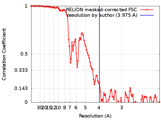
| Method | helical reconstruction / cryo EM / Resolution: 3.975 Å | ||||||||||||||||||
 Authors Authors | Iadanza MG / Ranson NA | ||||||||||||||||||
| Funding support |  United Kingdom, United Kingdom,  United States, 5 items United States, 5 items
| ||||||||||||||||||
 Citation Citation | Journal: Nat Commun / Year: 2018 Title: The structure of a β-microglobulin fibril suggests a molecular basis for its amyloid polymorphism. Authors: Matthew G Iadanza / Robert Silvers / Joshua Boardman / Hugh I Smith / Theodoros K Karamanos / Galia T Debelouchina / Yongchao Su / Robert G Griffin / Neil A Ranson / Sheena E Radford / Abstract: All amyloid fibrils contain a cross-β fold. How this structure differs in fibrils formed from proteins associated with different diseases remains unclear. Here, we combine cryo-EM and MAS-NMR to ...All amyloid fibrils contain a cross-β fold. How this structure differs in fibrils formed from proteins associated with different diseases remains unclear. Here, we combine cryo-EM and MAS-NMR to determine the structure of an amyloid fibril formed in vitro from β-microglobulin (βm), the culprit protein of dialysis-related amyloidosis. The fibril is composed of two identical protofilaments assembled from subunits that do not share βm's native tertiary fold, but are formed from similar β-strands. The fibrils share motifs with other amyloid fibrils, but also contain unique features including π-stacking interactions perpendicular to the fibril axis and an intramolecular disulfide that stabilises the subunit fold. We also describe a structural model for a second fibril morphology and show that it is built from the same subunit fold. The results provide insights into the mechanisms of fibril formation and the commonalities and differences within the amyloid fold in different protein sequences. | ||||||||||||||||||
| History |
|
- Structure visualization
| Structure viewer | EM map: SurfViewMolmilJmol/JSmol |
|---|---|
| Supplemental images |
- Downloads & links
-EMDB archive
| Map data | emd_0014.map.gz | 5.3 MB | EMDB map data format | |
|---|---|---|---|---|
| Header (meta data) | emd-0014-v30.xmlemd-0014.xml | 14.6 KB 14.6 KB | Display Display | EMDB header |
| FSC (resolution estimation) | emd_0014_fsc.xml | 10.7 KB | Display | FSC data file |
| Images |  emd_0014.png emd_0014.png | 163.4 KB | ||
| Filedesc metadata | emd-0014.cif.gz | 5.8 KB | ||
| Archive directory |  http://ftp.pdbj.org/pub/emdb/structures/EMD-0014ftp://ftp.pdbj.org/pub/emdb/structures/EMD-0014 http://ftp.pdbj.org/pub/emdb/structures/EMD-0014ftp://ftp.pdbj.org/pub/emdb/structures/EMD-0014 | HTTPS FTP |
-Related structure data
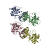
| Related structure data |  6gk3MC 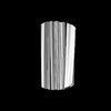 0021C M: atomic model generated by this map C: citing same article ( |
|---|---|
| Similar structure data | |
| EM raw data | EMPIAR-10207 (Title: Beta-2-microglobulin fibrils with multiple polymorphs formed at pH 2 Data size: 294.3 Data #1: Aligned, dose-weighted micrographs [micrographs - single frame]) |


-Links
| EMDB pages | EMDB (EBI/PDBe) / EMDataResource |
|---|---|
| Related items in Molecule of the Month |


-Map
| File | Download / File: emd_0014.map.gz / Format: CCP4 / Size: 103 MB / Type: IMAGE STORED AS FLOATING POINT NUMBER (4 BYTES) | ||||||||||||||||||||||||||||||||||||
|---|---|---|---|---|---|---|---|---|---|---|---|---|---|---|---|---|---|---|---|---|---|---|---|---|---|---|---|---|---|---|---|---|---|---|---|---|---|
| Annotation | Beta-2-microglobulin amyloid fibril - two protofilamets | ||||||||||||||||||||||||||||||||||||
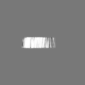
| Projections & slices | Image control
Images are generated by Spider. | ||||||||||||||||||||||||||||||||||||
| Voxel size | X=Y=Z: 1.06 Å | ||||||||||||||||||||||||||||||||||||
| Density |
| ||||||||||||||||||||||||||||||||||||
| Symmetry | Space group: 1 | ||||||||||||||||||||||||||||||||||||
| Details | EMDB XML:
|
 Z (Sec.)
 Y (Row.)
 X (Col.)


-Supplemental data
- Sample components
-Entire : two protofilament beta-2-microglobulin amyloid fibril
| Entire | Name: two protofilament beta-2-microglobulin amyloid fibril |
|---|---|
| Components |
|
-Supramolecule #1: two protofilament beta-2-microglobulin amyloid fibril
| Supramolecule | Name: two protofilament beta-2-microglobulin amyloid fibril / type: complex / ID: 1 / Parent: 0 / Macromolecule list: all |
|---|---|
| Source (natural) | Organism: Homo sapiens (human) |
| Molecular weight | Theoretical: 53.3 kDa/nm |
-Macromolecule #1: Beta-2-microglobulin
| Macromolecule | Name: Beta-2-microglobulin / type: protein_or_peptide / ID: 1 / Number of copies: 8 / Enantiomer: LEVO |
|---|---|
| Source (natural) | Organism: Homo sapiens (human) |
| Molecular weight | Theoretical: 7.635446 KDa |
| Recombinant expression | Organism:  |
| Sequence | String: FLNCYVSGFH PSDIEVDLLK NGERIEKVEH SDLSFSKDWS FYLLYYTEFT PTEKDEYACR VNHV UniProtKB: Beta-2-microglobulin |
-Experimental details
-Structure determination
| Method | cryo EM |
|---|---|
 Processing Processing | helical reconstruction |
| Aggregation state | filament |
-Sample preparation
| Concentration | 0.025 mg/mL | ||||||||||||
|---|---|---|---|---|---|---|---|---|---|---|---|---|---|
| Buffer | pH: 2.5 Component:
| ||||||||||||
| Grid | Model: Quantifoil R3.5/1 / Material: COPPER / Mesh: 400 / Support film - Material: CARBON / Support film - topology: HOLEY / Pretreatment - Type: PLASMA CLEANING / Pretreatment - Time: 60 sec. | ||||||||||||
| Vitrification | Cryogen name: ETHANE / Chamber humidity: 80 % / Chamber temperature: 281.15 K / Instrument: FEI VITROBOT MARK II | ||||||||||||
| Details | Quiescent growth at 0.25 mg/ml for 5 weeks, diluted 10x with buffer |
- Electron microscopy
| Microscope | FEI TITAN KRIOS |
|---|---|
| Image recording | Film or detector model: GATAN K2 SUMMIT (4k x 4k) / Detector mode: COUNTING / Digitization - Frames/image: 3-40 / Number grids imaged: 1 / Number real images: 5549 / Average exposure time: 10.0 sec. / Average electron dose: 38.5 e/Å2 |
| Electron beam | Acceleration voltage: 300 kV / Electron source:  FIELD EMISSION GUN FIELD EMISSION GUN |
| Electron optics | C2 aperture diameter: 100.0 µm / Illumination mode: FLOOD BEAM / Imaging mode: BRIGHT FIELD / Cs: 2.7 mm / Nominal defocus max: 0.0032500000000000003 µm / Nominal defocus min: 0.00175 µm / Nominal magnification: 130000 |
| Sample stage | Specimen holder model: FEI TITAN KRIOS AUTOGRID HOLDER / Cooling holder cryogen: NITROGEN |
| Experimental equipment |  Model: Titan Krios / Image courtesy: FEI Company |
+Image processing
-Atomic model buiding 1
| Refinement | Space: REAL / Protocol: OTHER |
|---|---|
| Output model | PDB-6gk3: |
