 Movie
Movie Controller

[English] 日本語
 Yorodumi
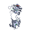
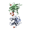
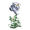
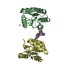
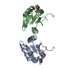
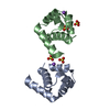
- PDB-1lxd: CRYSTAL STRUCTURE OF THE RAS INTERACTING DOMAIN OF RALGDS, A GUAN... -
+ Open data

- Basic information
| Entry | Database: PDB / ID: 1lxd | ||||||
|---|---|---|---|---|---|---|---|
| Title | CRYSTAL STRUCTURE OF THE RAS INTERACTING DOMAIN OF RALGDS, A GUANINE NUCLEOTIDE DISSOCIATION STIMULATOR OF RAL PROTEIN | ||||||
 Components Components | RALGDSB | ||||||
 Keywords Keywords | PHOSPHORYLATION / RALGDS / RAS BINDING / UBIQUITIN FOLD / CDC25 FAMILY / SIGNAL TRANSDUCTION / CROSS-TALK | ||||||
| Function / homology |  Function and homology information Function and homology informationp38MAPK events / GTPase regulator activity / RAF/MAP kinase cascade / small GTPase-mediated signal transduction / brush border / guanyl-nucleotide exchange factor activity / Ras protein signal transduction / nucleus / plasma membrane / cytosol Similarity search - Function | ||||||
| Biological species |  | ||||||
| Method |  X-RAY DIFFRACTION / SYNCHROTRON / MAD / Resolution: 2.4 Å X-RAY DIFFRACTION / SYNCHROTRON / MAD / Resolution: 2.4 Å | ||||||
 Authors Authors | Huang, L. / Weng, X.W. / Hofer, F. / Martin, G.S. / Kim, S.H. | ||||||
 Citation Citation | Journal: Nat.Struct.Biol. / Year: 1997 Title: Three-dimensional structure of the Ras-interacting domain of RalGDS. Authors: Huang, L. / Weng, X. / Hofer, F. / Martin, G.S. / Kim, S.H. #1: Journal: Acta Crystallogr.,Sect.D / Year: 1996Title: Crystallization and Preliminary Crystallographic Analysis of the Ras Binding Domain of Ralgds, a Guanine Nucleotide Dissociation Stimulator of the Ral Protein Authors: Huang, L. / Jancarik, J. / Kim, S.-H. / Hofer, F. / Martin, G.S. #2: Journal: Proc.Natl.Acad.Sci.USA / Year: 1994Title: Activated Ras Interacts with the Ral Guanine Nucleotide Dissociation Stimulator Authors: Hofer, F. / Fields, S. / Schneider, C. / Martin, G.S. | ||||||
| History |
|
- Structure visualization
| Structure viewer | Molecule: MolmilJmol/JSmol |
|---|
- Downloads & links
-Download
| PDBx/mmCIF format | 1lxd.cif.gz | 59.4 KB | Display | PDBx/mmCIF format |
|---|---|---|---|---|
| PDB format | pdb1lxd.ent.gz | 43.1 KB | Display | PDB format |
| PDBx/mmJSON format | 1lxd.json.gz | Tree view | PDBx/mmJSON format | |
| Others |  Other downloads Other downloads |
-Validation report
| Arichive directory | https://data.pdbj.org/pub/pdb/validation_reports/lx/1lxdftp://data.pdbj.org/pub/pdb/validation_reports/lx/1lxd | HTTPS FTP |
|---|
-Related structure data
| Similar structure data |
|---|


-Links
 PDBj
PDBj


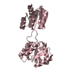
- Assembly
| Deposited unit | 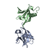
| ||||||||
|---|---|---|---|---|---|---|---|---|---|
| 1 |
| ||||||||
| Unit cell |
| ||||||||
| Noncrystallographic symmetry (NCS) | NCS oper: (Code: given Matrix: (-0.219734, -0.826478, -0.518316), Vector: |
-Components
| #1: Protein | Mass: 11350.771 Da / Num. of mol.: 2 / Fragment: C-TERMINAL DOMAIN WHICH BINDS TO ACTIVE RAS Mutation: N-TERMINAL GS INHERITED FROM THE LINKER SEQUENCE OF THE CLONING VECTOR Source method: isolated from a genetically manipulated source Source: (gene. exp.) Gene (production host): C-TERMINAL DOMAIN OF RALGDS FUSED TO GLUTATHIONINE S TRANSFERASE Production host:  #2: Water | ChemComp-HOH / |  Mass: 18.015 Da / Num. of mol.: 58 / Source method: isolated from a natural source / Formula: H2O Mass: 18.015 Da / Num. of mol.: 58 / Source method: isolated from a natural source / Formula: H2OHas protein modification | Y | Sequence details | TERMINAL RESIDUE LYS 100 IN THE PDB FILE IS LYS 864 IN THE RAT RALGDS SEQUENCE. | |
|---|
-Experimental details
-Experiment
| Experiment | Method: X-RAY DIFFRACTION / Number of used crystals: 1 |
|---|
- Sample preparation
| Crystal | Density Matthews: 1.8 Å3/Da / Density % sol: 30 % Description: THIS STRUCTURE WAS SOLVED USING THE MAD DATA ON THE SELENOMETHIONINE MUTANT OF THE PROTEIN AT X4A NSLS. BUT THE STRUCTURE WAS REFINED AGAINST THE NATIVE DATA ABOVE. | |||||||||||||||||||||||||||||||||||||||||||||||||||||||
|---|---|---|---|---|---|---|---|---|---|---|---|---|---|---|---|---|---|---|---|---|---|---|---|---|---|---|---|---|---|---|---|---|---|---|---|---|---|---|---|---|---|---|---|---|---|---|---|---|---|---|---|---|---|---|---|---|
| Crystal grow | pH: 8.5 Details: PROTEIN WAS CRYSTALLIZED FROM 20% PEG 8000, 0.1 M TRIS PH 8.5, AND 0.2 M CALCIUM ACETATE. | |||||||||||||||||||||||||||||||||||||||||||||||||||||||
| Crystal grow | *PLUS Method: vapor diffusion | |||||||||||||||||||||||||||||||||||||||||||||||||||||||
| Components of the solutions | *PLUS
|
-Data collection
| Diffraction | Mean temperature: 100 K |
|---|---|
| Diffraction source | Source: SYNCHROTRON / Site: NSLS  / Beamline: X12B / Wavelength: 1.008 / Beamline: X12B / Wavelength: 1.008 |
| Detector | Type: MARRESEARCH / Detector: IMAGE PLATE / Date: Nov 10, 1996 / Details: MIRRORS |
| Radiation | Monochromator: SI(111) / Monochromatic (M) / Laue (L): M / Scattering type: x-ray |
| Radiation wavelength | Wavelength: 1.008 Å / Relative weight: 1 |
| Reflection | Resolution: 2.4→10 Å / Num. obs: 6485 / % possible obs: 99.3 % / Observed criterion σ(I): 0 / Redundancy: 4 % / Biso Wilson estimate: 20 Å2 / Rsym value: 0.09 / Net I/σ(I): 16.7 |
| Reflection shell | Resolution: 2.4→2.48 Å / Redundancy: 4 % / Mean I/σ(I) obs: 5.92 / Rsym value: 0.251 / % possible all: 99.5 |
| Reflection | *PLUS Rmerge(I) obs: 0.09 |
- Processing
| Software |
| ||||||||||||||||||||||||||||||||||||||||||||||||||||||||||||
|---|---|---|---|---|---|---|---|---|---|---|---|---|---|---|---|---|---|---|---|---|---|---|---|---|---|---|---|---|---|---|---|---|---|---|---|---|---|---|---|---|---|---|---|---|---|---|---|---|---|---|---|---|---|---|---|---|---|---|---|---|---|
| Refinement | Method to determine structure: MAD / Resolution: 2.4→6 Å / Rfactor Rfree error: 0.04 / Data cutoff high absF: 15000 / Data cutoff low absF: 0 / Cross valid method: THROUGHOUT / σ(F): 2 / Details: H19A.PEP IS THE PEPTIDE BOND FIL
| ||||||||||||||||||||||||||||||||||||||||||||||||||||||||||||
| Displacement parameters | Biso mean: 15 Å2 | ||||||||||||||||||||||||||||||||||||||||||||||||||||||||||||
| Refine analyze | Luzzati d res low obs: 6 Å | ||||||||||||||||||||||||||||||||||||||||||||||||||||||||||||
| Refinement step | Cycle: LAST / Resolution: 2.4→6 Å
| ||||||||||||||||||||||||||||||||||||||||||||||||||||||||||||
| Refine LS restraints |
| ||||||||||||||||||||||||||||||||||||||||||||||||||||||||||||
| Refine LS restraints NCS | NCS model details: NO RESTRAINTS. | ||||||||||||||||||||||||||||||||||||||||||||||||||||||||||||
| LS refinement shell | Resolution: 2.4→2.48 Å / Rfactor Rfree error: 0.045 / Total num. of bins used: 10
| ||||||||||||||||||||||||||||||||||||||||||||||||||||||||||||
| Xplor file |
| ||||||||||||||||||||||||||||||||||||||||||||||||||||||||||||
| Software | *PLUS Name: X-PLOR / Version: 3.85 / Classification: refinement | ||||||||||||||||||||||||||||||||||||||||||||||||||||||||||||
| LS refinement shell | *PLUS Rfactor Rfree: 0.31 |
