 Movie
Movie Controller


+ Open data

- Basic information
| Entry | Database: PDB / ID: 1b5e | ||||||
|---|---|---|---|---|---|---|---|
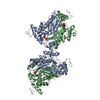
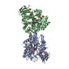
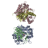
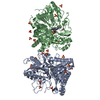
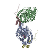
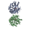
| Title | DCMP HYDROXYMETHYLASE FROM T4 | ||||||
 Components Components | PROTEIN (DEOXYCYTIDYLATE HYDROXYMETHYLASE) | ||||||
 Keywords Keywords | TRANSFERASE / HYDROXYMETHYLASE / DNTP SYNTHESIZING COMPLEX | ||||||
| Function / homology |  Function and homology information Function and homology informationdeoxycytidylate 5-hydroxymethyltransferase / deoxycytidylate 5-hydroxymethyltransferase activity Similarity search - Function | ||||||
| Biological species |  Enterobacteria phage T4 (virus) Enterobacteria phage T4 (virus) | ||||||
| Method |  X-RAY DIFFRACTION / SYNCHROTRON / MIR / Resolution: 1.6 Å X-RAY DIFFRACTION / SYNCHROTRON / MIR / Resolution: 1.6 Å | ||||||
 Authors Authors | Song, H.K. / Sohn, S.H. / Suh, S.W. | ||||||
 Citation Citation | Journal: EMBO J. / Year: 1999 Title: Crystal structure of deoxycytidylate hydroxymethylase from bacteriophage T4, a component of the deoxyribonucleoside triphosphate-synthesizing complex. Authors: Song, H.K. / Sohn, S.H. / Suh, S.W. | ||||||
| History |
|
- Structure visualization
| Structure viewer | Molecule: MolmilJmol/JSmol |
|---|
- Downloads & links
-Download
| PDBx/mmCIF format | 1b5e.cif.gz | 111 KB | Display | PDBx/mmCIF format |
|---|---|---|---|---|
| PDB format | pdb1b5e.ent.gz | 86.2 KB | Display | PDB format |
| PDBx/mmJSON format | 1b5e.json.gz | Tree view | PDBx/mmJSON format | |
| Others |  Other downloads Other downloads |
-Validation report
| Arichive directory | https://data.pdbj.org/pub/pdb/validation_reports/b5/1b5eftp://data.pdbj.org/pub/pdb/validation_reports/b5/1b5e | HTTPS FTP |
|---|
-Related structure data


-Links
 PDBj
PDBj- Assembly
| Deposited unit | 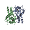
| ||||||||
|---|---|---|---|---|---|---|---|---|---|
| 1 | 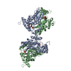
| ||||||||
| 2 |
| ||||||||
| Unit cell |
| ||||||||
| Noncrystallographic symmetry (NCS) | NCS oper: (Code: given Matrix: (0.630999, 0.000917, 0.775783), Vector: |
-Components
| #1: Protein | Mass: 28523.229 Da / Num. of mol.: 2 / Source method: isolated from a natural source / Source: (natural) Enterobacteria phage T4 (virus) / Genus: T4-like viruses / Species: Enterobacteria phage T4 sensu latoReferences: UniProt: P08773, deoxycytidylate 5-hydroxymethyltransferase #2: Chemical |   Mass: 307.197 Da / Num. of mol.: 2 / Source method: obtained synthetically / Formula: C9H14N3O7P Mass: 307.197 Da / Num. of mol.: 2 / Source method: obtained synthetically / Formula: C9H14N3O7P#3: Water | ChemComp-HOH / |  Mass: 18.015 Da / Num. of mol.: 158 / Source method: isolated from a natural source / Formula: H2O Mass: 18.015 Da / Num. of mol.: 158 / Source method: isolated from a natural source / Formula: H2O |
|---|
-Experimental details
-Experiment
| Experiment | Method: X-RAY DIFFRACTION / Number of used crystals: 2 |
|---|
- Sample preparation
| Crystal | Density Matthews: 2.65 Å3/Da / Density % sol: 54 % Description: DATA WERE COLLECTED USING THE WEISSENBERG METHOD. | ||||||||||||||||||||||||||||||
|---|---|---|---|---|---|---|---|---|---|---|---|---|---|---|---|---|---|---|---|---|---|---|---|---|---|---|---|---|---|---|---|
| Crystal grow | pH: 8 / Details: pH 8.0 | ||||||||||||||||||||||||||||||
| Crystal | *PLUS | ||||||||||||||||||||||||||||||
| Crystal grow | *PLUS Method: vapor diffusion, hanging drop / Details: Song, S.H., (1999) Acta Crystallogr. D55, 1061. / pH: 8.73 | ||||||||||||||||||||||||||||||
| Components of the solutions | *PLUS
|
-Data collection
| Diffraction | Mean temperature: 293 K |
|---|---|
| Diffraction source | Source: SYNCHROTRON / Site: Photon Factory  / Beamline: BL-6A / Wavelength: 1 / Beamline: BL-6A / Wavelength: 1 |
| Detector | Type: FUJI / Detector: IMAGE PLATE / Date: Apr 15, 1998 |
| Radiation | Protocol: SINGLE WAVELENGTH / Monochromatic (M) / Laue (L): M / Scattering type: x-ray |
| Radiation wavelength | Wavelength: 1 Å / Relative weight: 1 |
| Reflection | Resolution: 1.6→50 Å / Num. obs: 80033 / % possible obs: 97.3 % / Redundancy: 6.4 % / Rsym value: 0.066 |
- Processing
| Software |
| ||||||||||||||||||||||||||||||||||||||||||||||||||||||||||||
|---|---|---|---|---|---|---|---|---|---|---|---|---|---|---|---|---|---|---|---|---|---|---|---|---|---|---|---|---|---|---|---|---|---|---|---|---|---|---|---|---|---|---|---|---|---|---|---|---|---|---|---|---|---|---|---|---|---|---|---|---|---|
| Refinement | Method to determine structure: MIR / Resolution: 1.6→8 Å / Cross valid method: THROUGHOUT / σ(F): 0
| ||||||||||||||||||||||||||||||||||||||||||||||||||||||||||||
| Refinement step | Cycle: LAST / Resolution: 1.6→8 Å
| ||||||||||||||||||||||||||||||||||||||||||||||||||||||||||||
| Refine LS restraints |
| ||||||||||||||||||||||||||||||||||||||||||||||||||||||||||||
| Xplor file | Serial no: 3 / Param file: PARHCSDX.PRO / Topol file: TOPHCSDX.PRO | ||||||||||||||||||||||||||||||||||||||||||||||||||||||||||||
| Software | *PLUS Name: X-PLOR / Version: 3.843 / Classification: refinement | ||||||||||||||||||||||||||||||||||||||||||||||||||||||||||||
| Refinement | *PLUS σ(F): 0 / % reflection Rfree: 5 % | ||||||||||||||||||||||||||||||||||||||||||||||||||||||||||||
| Solvent computation | *PLUS | ||||||||||||||||||||||||||||||||||||||||||||||||||||||||||||
| Displacement parameters | *PLUS |
