protein C (activated) / positive regulation of establishment of endothelial barrier / negative regulation of coagulation / negative regulation of blood coagulation / Gamma-carboxylation of protein precursors / Transport of gamma-carboxylated protein precursors from the endoplasmic reticulum to the Golgi apparatus / Common Pathway of Fibrin Clot Formation / Removal of aminoterminal propeptides from gamma-carboxylated proteins / Intrinsic Pathway of Fibrin Clot Formation / Cell surface interactions at the vascular wall ...protein C (activated) / positive regulation of establishment of endothelial barrier / negative regulation of coagulation / negative regulation of blood coagulation / Gamma-carboxylation of protein precursors / Transport of gamma-carboxylated protein precursors from the endoplasmic reticulum to the Golgi apparatus / Common Pathway of Fibrin Clot Formation / Removal of aminoterminal propeptides from gamma-carboxylated proteins / Intrinsic Pathway of Fibrin Clot Formation / Cell surface interactions at the vascular wall / Post-translational protein phosphorylation / negative regulation of inflammatory response / Golgi lumen / Regulation of Insulin-like Growth Factor (IGF) transport and uptake by Insulin-like Growth Factor Binding Proteins (IGFBPs) / blood coagulation / endoplasmic reticulum lumen / serine-type endopeptidase activity / calcium ion binding / negative regulation of apoptotic process / Golgi apparatus / endoplasmic reticulum / proteolysis / extracellular space / extracellular region Similarity search - Function
Mass: 18.015 Da / Num. of mol.: 146 / Source method: isolated from a natural source / Formula: H2O
Nonpolymer details
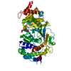
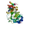
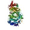
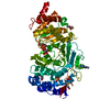
THE UNBOUND FORM OF THE INHIBITOR IS D-PHE-PRO-ARG-CHLOROMETHYLKETONE. UPON REACTION WITH PROTEIN ...THE UNBOUND FORM OF THE INHIBITOR IS D-PHE-PRO-ARG-CHLOROMETHYLKETONE. UPON REACTION WITH PROTEIN IT FORMS TWO COVALENT BONDS: 1) A COVALENT BOND TO SER 195 FORMING A HEMIKETAL AR7 AND 2) A COVALENT BOND TO NE2 OF HIS 57
Sequence details
THE CHYMOTRYPSIN NUMBERING (RATHER THAN SEQUENTIAL) SYSTEM IS USED HERE, BASED ON THE TOPOLOGICAL ...THE CHYMOTRYPSIN NUMBERING (RATHER THAN SEQUENTIAL) SYSTEM IS USED HERE, BASED ON THE TOPOLOGICAL ALIGNMENT WITH THE STRUCTURE OF CHYMOTRYPSIN (W.BODE ET AL., 1989, EMBO J.8, 3467-3475).
-
Experimental details
-
Experiment
Experiment
Method: X-RAY DIFFRACTION / Number of used crystals: 1
-
Sample preparation
Crystal
Density Matthews: 3.14 Å3/Da / Density % sol: 61 %
Method to determine structure: MOLECULAR REPLACEMENT Starting model: FACTOR XA POLY-ALA Resolution: 2.8→8 Å / σ(F): 2 Details: THE FOLLOWING SEGMENTS ARE DISORDERED AND ARE MODELED STEREOCHEMICALLY TO PRESERVE CHAIN CONTINUITY: C 60 - C 62, C 148 - C 151, L 70 - L 77. HIS L 107 IS IN A TIGHT TURN AND HAS TORSION ...Details: THE FOLLOWING SEGMENTS ARE DISORDERED AND ARE MODELED STEREOCHEMICALLY TO PRESERVE CHAIN CONTINUITY: C 60 - C 62, C 148 - C 151, L 70 - L 77. HIS L 107 IS IN A TIGHT TURN AND HAS TORSION ANGLES OUTSIDE THE EXPECTED RAMACHANDRAN REGIONS. THE STRAIN IS STABILIZED BY A HYDROGEN BOND TO ASN L 102. RESIDUE BHD L 71 HAS BEEN SHOWN TO BE BETA-HYDROXYLATED ALTHOUGH IT IS IN A DISORDERED SEGMENT OF THIS STRUCTURE. THE HYDROXYL GROUP IS NOT VISIBLE.
Rfactor
Num. reflection
Rwork
0.184
-
obs
-
11715
Displacement parameters
Biso mean: 26.7 Å2
Refinement step
Cycle: LAST / Resolution: 2.8→8 Å
Protein
Nucleic acid
Ligand
Solvent
Total
Num. atoms
2635
0
30
146
2811
Refine LS restraints
Refine-ID
Type
Dev ideal
Dev ideal target
X-RAY DIFFRACTION
p_bond_d
0.012
0.02
X-RAY DIFFRACTION
p_angle_d
2.072
0.04
X-RAY DIFFRACTION
p_angle_deg
X-RAY DIFFRACTION
p_planar_d
0.05
X-RAY DIFFRACTION
p_hb_or_metal_coord
X-RAY DIFFRACTION
p_mcbond_it
1.5
X-RAY DIFFRACTION
p_mcangle_it
2
X-RAY DIFFRACTION
p_scbond_it
2
X-RAY DIFFRACTION
p_scangle_it
2.5
X-RAY DIFFRACTION
p_plane_restr
0.01
X-RAY DIFFRACTION
p_chiral_restr
0.1
X-RAY DIFFRACTION
p_singtor_nbd
0.2
X-RAY DIFFRACTION
p_multtor_nbd
0.2
X-RAY DIFFRACTION
p_xhyhbond_nbd
X-RAY DIFFRACTION
p_xyhbond_nbd
0.2
X-RAY DIFFRACTION
p_planar_tor
10
X-RAY DIFFRACTION
p_staggered_tor
15
X-RAY DIFFRACTION
p_orthonormal_tor
X-RAY DIFFRACTION
p_transverse_tor
45
X-RAY DIFFRACTION
p_special_tor
Software
*PLUS
Name: PROLSQ / Classification: refinement
Refinement
*PLUS
Rfactor obs: 0.184
Solvent computation
*PLUS
Displacement parameters
*PLUS
+
About Yorodumi
-
News
-
Feb 9, 2022. New format data for meta-information of EMDB entries
New format data for meta-information of EMDB entries
Version 3 of the EMDB header file is now the official format.
The previous official version 1.9 will be removed from the archive.
In the structure databanks used in Yorodumi, some data are registered as the other names, "COVID-19 virus" and "2019-nCoV". Here are the details of the virus and the list of structure data.
Jan 31, 2019. EMDB accession codes are about to change! (news from PDBe EMDB page)
EMDB accession codes are about to change! (news from PDBe EMDB page)
The allocation of 4 digits for EMDB accession codes will soon come to an end. Whilst these codes will remain in use, new EMDB accession codes will include an additional digit and will expand incrementally as the available range of codes is exhausted. The current 4-digit format prefixed with “EMD-” (i.e. EMD-XXXX) will advance to a 5-digit format (i.e. EMD-XXXXX), and so on. It is currently estimated that the 4-digit codes will be depleted around Spring 2019, at which point the 5-digit format will come into force.
The EM Navigator/Yorodumi systems omit the EMD- prefix.
Related info.:Q: What is EMD? / ID/Accession-code notation in Yorodumi/EM Navigator
Yorodumi is a browser for structure data from EMDB, PDB, SASBDB, etc.
This page is also the successor to EM Navigator detail page, and also detail information page/front-end page for Omokage search.
The word "yorodu" (or yorozu) is an old Japanese word meaning "ten thousand". "mi" (miru) is to see.
Related info.:EMDB / PDB / SASBDB / Comparison of 3 databanks / Yorodumi Search / Aug 31, 2016. New EM Navigator & Yorodumi / Yorodumi Papers / Jmol/JSmol / Function and homology information / Changes in new EM Navigator and Yorodumi
 Movie
Movie Controller


 Open data
 Basic information
 Components
 Keywords
 Function and homology information
 Homo sapiens (human)
 X-RAY DIFFRACTION /
  Authors
 Citation
 Structure visualization
 Downloads & links
 Other downloads
 PDBj
PDBj


 Assembly

 Type: peptide-like, Peptide-like / Class: Inhibitor / Mass: 453.986 Da / Num. of mol.: 1 / Source method: obtained synthetically / Formula: C21H34ClN6O3 / Details: MAI IS DEOXO-METHYLARGININE SYNTHETIC PEPTIDE / References: D-Phe-Pro-Arg-CH2Cl
 Mass: 18.015 Da / Num. of mol.: 146 / Source method: isolated from a natural source / Formula: H2O
 Sample preparation
 Processing