+ Open data
- Basic information
| Entry | Database: EMDB / ID: EMD-9196 | |||||||||
|---|---|---|---|---|---|---|---|---|---|---|
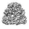
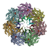
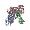
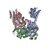
| Title | ADP-bound human mitochondrial Hsp60-Hsp10 half-football complex | |||||||||
 Map data Map data | ADP-bound mHsp60-mHsp10 half-football complex | |||||||||
 Sample Sample |
| |||||||||
 Keywords Keywords | Complex / ADP / Half-football / CHAPERONE | |||||||||
| Function / homology |  Function and homology information Function and homology informationcoated vesicle / isotype switching to IgG isotypes / mitochondrial unfolded protein response / TFAP2A acts as a transcriptional repressor during retinoic acid induced cell differentiation / apolipoprotein A-I binding / lipopolysaccharide receptor complex / protein import into mitochondrial intermembrane space / high-density lipoprotein particle binding / migrasome / cysteine-type endopeptidase activator activity ...coated vesicle / isotype switching to IgG isotypes / mitochondrial unfolded protein response / TFAP2A acts as a transcriptional repressor during retinoic acid induced cell differentiation / apolipoprotein A-I binding / lipopolysaccharide receptor complex / protein import into mitochondrial intermembrane space / high-density lipoprotein particle binding / migrasome / cysteine-type endopeptidase activator activity / positive regulation of T cell mediated immune response to tumor cell / chaperonin ATPase / Mitochondrial protein import / positive regulation of macrophage activation / negative regulation of execution phase of apoptosis / cellular response to interleukin-7 / MyD88-dependent toll-like receptor signaling pathway / 'de novo' protein folding / biological process involved in interaction with symbiont / sperm plasma membrane / apoptotic mitochondrial changes / : / B cell activation / B cell proliferation / positive regulation of interferon-alpha production / DNA replication origin binding / positive regulation of interleukin-10 production / apolipoprotein binding / RHOG GTPase cycle / positive regulation of execution phase of apoptosis / response to unfolded protein / Mitochondrial unfolded protein response (UPRmt) / isomerase activity / chaperone-mediated protein complex assembly / sperm midpiece / clathrin-coated pit / positive regulation of interleukin-12 production / protein folding chaperone / Mitochondrial protein degradation / intrinsic apoptotic signaling pathway / response to cold / secretory granule / T cell activation / protein maturation / ATP-dependent protein folding chaperone / lipopolysaccharide binding / positive regulation of T cell activation / positive regulation of interleukin-6 production / positive regulation of type II interferon production / osteoblast differentiation / p53 binding / unfolded protein binding / protein folding / single-stranded DNA binding / double-stranded RNA binding / protein-folding chaperone binding / protein refolding / early endosome / mitochondrial inner membrane / protein stabilization / mitochondrial matrix / ubiquitin protein ligase binding / negative regulation of apoptotic process / enzyme binding / cell surface / ATP hydrolysis activity / protein-containing complex / mitochondrion / extracellular space / RNA binding / extracellular exosome / ATP binding / metal ion binding / membrane / plasma membrane / cytosol / cytoplasm Similarity search - Function | |||||||||
| Biological species |  Homo sapiens (human) Homo sapiens (human) | |||||||||
| Method | single particle reconstruction / cryo EM / Resolution: 3.82 Å | |||||||||
 Authors Authors | Gomez-Llorente Y / Jebara F / Patra M | |||||||||
| Funding support |  United States, 1 items United States, 1 items
| |||||||||
 Citation Citation |  Journal: Nat Commun / Year: 2020 Journal: Nat Commun / Year: 2020Title: Structural basis for active single and double ring complexes in human mitochondrial Hsp60-Hsp10 chaperonin. Authors: Yacob Gomez-Llorente / Fady Jebara / Malay Patra / Radhika Malik / Shahar Nisemblat / Orna Chomsky-Hecht / Avital Parnas / Abdussalam Azem / Joel A Hirsch / Iban Ubarretxena-Belandia /    Abstract: mHsp60-mHsp10 assists the folding of mitochondrial matrix proteins without the negative ATP binding inter-ring cooperativity of GroEL-GroES. Here we report the crystal structure of an ATP (ADP:BeF- ...mHsp60-mHsp10 assists the folding of mitochondrial matrix proteins without the negative ATP binding inter-ring cooperativity of GroEL-GroES. Here we report the crystal structure of an ATP (ADP:BeF-bound) ground-state mimic double-ring mHsp60-(mHsp10) football complex, and the cryo-EM structures of the ADP-bound successor mHsp60-(mHsp10) complex, and a single-ring mHsp60-mHsp10 half-football. The structures explain the nucleotide dependence of mHsp60 ring formation, and reveal an inter-ring nucleotide symmetry consistent with the absence of negative cooperativity. In the ground-state a two-fold symmetric H-bond and a salt bridge stitch the double-rings together, whereas only the H-bond remains as the equatorial gap increases in an ADP football poised to split into half-footballs. Refolding assays demonstrate obligate single- and double-ring mHsp60 variants are active, and complementation analysis in bacteria shows the single-ring variant is as efficient as wild-type mHsp60. Our work provides a structural basis for active single- and double-ring complexes coexisting in the mHsp60-mHsp10 chaperonin reaction cycle. | |||||||||
| History |
|
- Structure visualization
| Movie |
 Movie viewer Movie viewer |
|---|---|
| Structure viewer | EM map:  SurfView SurfView Molmil Molmil Jmol/JSmol Jmol/JSmol |
| Supplemental images |
- Downloads & links
-EMDB archive
| Map data |  emd_9196.map.gz emd_9196.map.gz | 156.2 MB |  EMDB map data format EMDB map data format | |
|---|---|---|---|---|
| Header (meta data) |  emd-9196-v30.xml emd-9196-v30.xml emd-9196.xml emd-9196.xml | 13.1 KB 13.1 KB | Display Display |  EMDB header EMDB header |
| Images |  emd_9196.png emd_9196.png | 79.9 KB | ||
| Filedesc metadata |  emd-9196.cif.gz emd-9196.cif.gz | 6.1 KB | ||
| Archive directory |  http://ftp.pdbj.org/pub/emdb/structures/EMD-9196 http://ftp.pdbj.org/pub/emdb/structures/EMD-9196 ftp://ftp.pdbj.org/pub/emdb/structures/EMD-9196 ftp://ftp.pdbj.org/pub/emdb/structures/EMD-9196 | HTTPS FTP |
-Validation report
| Summary document |  emd_9196_validation.pdf.gz emd_9196_validation.pdf.gz | 562.1 KB | Display |  EMDB validaton report EMDB validaton report |
|---|---|---|---|---|
| Full document |  emd_9196_full_validation.pdf.gz emd_9196_full_validation.pdf.gz | 561.6 KB | Display | |
| Data in XML |  emd_9196_validation.xml.gz emd_9196_validation.xml.gz | 6.9 KB | Display | |
| Data in CIF |  emd_9196_validation.cif.gz emd_9196_validation.cif.gz | 7.9 KB | Display | |
| Arichive directory |  https://ftp.pdbj.org/pub/emdb/validation_reports/EMD-9196 https://ftp.pdbj.org/pub/emdb/validation_reports/EMD-9196 ftp://ftp.pdbj.org/pub/emdb/validation_reports/EMD-9196 ftp://ftp.pdbj.org/pub/emdb/validation_reports/EMD-9196 | HTTPS FTP |
-Related structure data
| Related structure data |  6mrdMC  9195C  6mrcC C: citing same article ( M: atomic model generated by this map |
|---|---|
| Similar structure data |
- Links
Links
| EMDB pages |  EMDB (EBI/PDBe) / EMDB (EBI/PDBe) /  EMDataResource EMDataResource |
|---|---|
| Related items in Molecule of the Month |
- Map
Map
| File |  Download / File: emd_9196.map.gz / Format: CCP4 / Size: 166.4 MB / Type: IMAGE STORED AS FLOATING POINT NUMBER (4 BYTES) Download / File: emd_9196.map.gz / Format: CCP4 / Size: 166.4 MB / Type: IMAGE STORED AS FLOATING POINT NUMBER (4 BYTES) | ||||||||||||||||||||||||||||||||||||||||||||||||||||||||||||||||||||
|---|---|---|---|---|---|---|---|---|---|---|---|---|---|---|---|---|---|---|---|---|---|---|---|---|---|---|---|---|---|---|---|---|---|---|---|---|---|---|---|---|---|---|---|---|---|---|---|---|---|---|---|---|---|---|---|---|---|---|---|---|---|---|---|---|---|---|---|---|---|
| Annotation | ADP-bound mHsp60-mHsp10 half-football complex | ||||||||||||||||||||||||||||||||||||||||||||||||||||||||||||||||||||
| Projections & slices | Image control
Images are generated by Spider. | ||||||||||||||||||||||||||||||||||||||||||||||||||||||||||||||||||||
| Voxel size | X=Y=Z: 1.07 Å | ||||||||||||||||||||||||||||||||||||||||||||||||||||||||||||||||||||
| Density |
| ||||||||||||||||||||||||||||||||||||||||||||||||||||||||||||||||||||
| Symmetry | Space group: 1 | ||||||||||||||||||||||||||||||||||||||||||||||||||||||||||||||||||||
| Details | EMDB XML:
CCP4 map header:
| ||||||||||||||||||||||||||||||||||||||||||||||||||||||||||||||||||||
-Supplemental data
- Sample components
-Entire : Half-football complex of Hsp60 with Hsp10 bound to ADP and Mg
| Entire | Name: Half-football complex of Hsp60 with Hsp10 bound to ADP and Mg |
|---|---|
| Components |
|
-Supramolecule #1: Half-football complex of Hsp60 with Hsp10 bound to ADP and Mg
| Supramolecule | Name: Half-football complex of Hsp60 with Hsp10 bound to ADP and Mg type: complex / ID: 1 / Parent: 0 / Macromolecule list: #1-#2 |
|---|---|
| Source (natural) | Organism:  Homo sapiens (human) Homo sapiens (human) |
| Molecular weight | Theoretical: 490 KDa |
-Macromolecule #1: 60 kDa heat shock protein, mitochondrial
| Macromolecule | Name: 60 kDa heat shock protein, mitochondrial / type: protein_or_peptide / ID: 1 / Number of copies: 7 / Enantiomer: LEVO / EC number: ec: 3.6.4.9 |
|---|---|
| Source (natural) | Organism:  Homo sapiens (human) Homo sapiens (human) |
| Molecular weight | Theoretical: 56.263559 KDa |
| Recombinant expression | Organism:  |
| Sequence | String: GSAKDVKFGA DARALMLQGV DLLADAVAVT MGPKGRTVII EQSWGSPKVT KDGVTVAKSI DLKDKYKNIG AKLVQDVANN TNEEAGDGT TTATVLARSI AKEGFEKISK GANPVEIRRG VMLAVDAVIA ELKKQSKPVT TPEEIAQVAT ISANGDKEIG N IISDAMKK ...String: GSAKDVKFGA DARALMLQGV DLLADAVAVT MGPKGRTVII EQSWGSPKVT KDGVTVAKSI DLKDKYKNIG AKLVQDVANN TNEEAGDGT TTATVLARSI AKEGFEKISK GANPVEIRRG VMLAVDAVIA ELKKQSKPVT TPEEIAQVAT ISANGDKEIG N IISDAMKK VGRKGVITVK DGKTLNDELE IIEGMKFDRG YISPYFINTS KGQKCEFQDA YVLLSEKKIS SIQSIVPALE IA NAHRKPL VIIAEDVDGE ALSTLVLNRL KVGLQVVAVK APGFGDNRKN QLKDMAIATG GAVFGEEGLT LNLEDVQPHD LGK VGEVIV TKDDAMLLKG KGDKAQIEKR IQEIIEQLDV TTSEYEKEKL NERLAKLSDG VAVLKVGGTS DVEVNEKKDR VTDA LNATR AAVEEGIVLG GGCALLRCIP ALDSLTPANE DQKIGIEIIK RTLKIPAMTI AKNAGVEGSL IVEKIMQSSS EVGYD AMAG DFVNMVEKGI IDPTKVVRTA LLDAAGVASL LTTAEVVVTE IPKE UniProtKB: 60 kDa heat shock protein, mitochondrial |
-Macromolecule #2: 10 kDa heat shock protein, mitochondrial
| Macromolecule | Name: 10 kDa heat shock protein, mitochondrial / type: protein_or_peptide / ID: 2 / Number of copies: 7 / Enantiomer: LEVO |
|---|---|
| Source (natural) | Organism:  Homo sapiens (human) Homo sapiens (human) |
| Molecular weight | Theoretical: 10.7444 KDa |
| Recombinant expression | Organism:  |
| Sequence | String: GQAFRKFLPL FDRVLVERSA AETVTKGGIM LPEKSQGKVL QATVVAVGSG SKGKGGEIQP VSVKVGDKVL LPEYGGTKVV LDDKDYFLF RDGDILGKYV D UniProtKB: 10 kDa heat shock protein, mitochondrial |
-Macromolecule #3: ADENOSINE-5'-DIPHOSPHATE
| Macromolecule | Name: ADENOSINE-5'-DIPHOSPHATE / type: ligand / ID: 3 / Number of copies: 7 / Formula: ADP |
|---|---|
| Molecular weight | Theoretical: 427.201 Da |
| Chemical component information |  ChemComp-ADP: |
-Macromolecule #4: MAGNESIUM ION
| Macromolecule | Name: MAGNESIUM ION / type: ligand / ID: 4 / Number of copies: 7 / Formula: MG |
|---|---|
| Molecular weight | Theoretical: 24.305 Da |
-Experimental details
-Structure determination
| Method | cryo EM |
|---|---|
 Processing Processing | single particle reconstruction |
| Aggregation state | particle |
- Sample preparation
| Buffer | pH: 7.7 |
|---|---|
| Grid | Support film - Material: CARBON / Support film - topology: LACEY / Details: unspecified |
| Vitrification | Cryogen name: ETHANE / Instrument: HOMEMADE PLUNGER |
- Electron microscopy
| Microscope | FEI TITAN KRIOS |
|---|---|
| Image recording | Film or detector model: GATAN K2 SUMMIT (4k x 4k) / Detector mode: COUNTING / Average exposure time: 10.0 sec. / Average electron dose: 63.0 e/Å2 |
| Electron beam | Acceleration voltage: 300 kV / Electron source:  FIELD EMISSION GUN FIELD EMISSION GUN |
| Electron optics | Illumination mode: FLOOD BEAM / Imaging mode: BRIGHT FIELD |
| Sample stage | Specimen holder model: FEI TITAN KRIOS AUTOGRID HOLDER / Cooling holder cryogen: NITROGEN |
| Experimental equipment |  Model: Titan Krios / Image courtesy: FEI Company |
- Image processing
| Startup model | Type of model: OTHER |
|---|---|
| Final reconstruction | Applied symmetry - Point group: C7 (7 fold cyclic) / Resolution.type: BY AUTHOR / Resolution: 3.82 Å / Resolution method: FSC 0.143 CUT-OFF / Software - Name: cryoSPARC / Number images used: 10972 |
| Initial angle assignment | Type: MAXIMUM LIKELIHOOD / Software - Name: RELION |
| Final angle assignment | Type: MAXIMUM LIKELIHOOD |
 Movie
Movie Controller


 Z (Sec.)
 Y (Row.)
 X (Col.)


