 Movie
Movie Controller


+ Open data

- Basic information
| Entry | Database: PDB / ID: 6mrc | ||||||||||||||||||||||||||||||||||||||||||
|---|---|---|---|---|---|---|---|---|---|---|---|---|---|---|---|---|---|---|---|---|---|---|---|---|---|---|---|---|---|---|---|---|---|---|---|---|---|---|---|---|---|---|---|
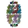
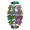
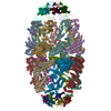
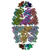
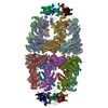
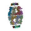
| Title | ADP-bound human mitochondrial Hsp60-Hsp10 football complex | ||||||||||||||||||||||||||||||||||||||||||
 Components Components |
| ||||||||||||||||||||||||||||||||||||||||||
 Keywords Keywords | CHAPERONE / Complex / ADP / Football | ||||||||||||||||||||||||||||||||||||||||||
| Function / homology |  Function and homology information Function and homology informationcoated vesicle / isotype switching to IgG isotypes / mitochondrial unfolded protein response / TFAP2A acts as a transcriptional repressor during retinoic acid induced cell differentiation / apolipoprotein A-I binding / lipopolysaccharide receptor complex / protein import into mitochondrial intermembrane space / high-density lipoprotein particle binding / migrasome / cysteine-type endopeptidase activator activity ...coated vesicle / isotype switching to IgG isotypes / mitochondrial unfolded protein response / TFAP2A acts as a transcriptional repressor during retinoic acid induced cell differentiation / apolipoprotein A-I binding / lipopolysaccharide receptor complex / protein import into mitochondrial intermembrane space / high-density lipoprotein particle binding / migrasome / cysteine-type endopeptidase activator activity / positive regulation of T cell mediated immune response to tumor cell / Mitochondrial protein import / positive regulation of macrophage activation / negative regulation of execution phase of apoptosis / chaperonin ATPase / cellular response to interleukin-7 / MyD88-dependent toll-like receptor signaling pathway / 'de novo' protein folding / sperm plasma membrane / biological process involved in interaction with symbiont / apoptotic mitochondrial changes / B cell proliferation / positive regulation of interferon-alpha production / B cell activation / positive regulation of interleukin-10 production / DNA replication origin binding / RHOG GTPase cycle / apolipoprotein binding / response to unfolded protein / Mitochondrial unfolded protein response (UPRmt) / positive regulation of execution phase of apoptosis / isomerase activity / chaperone-mediated protein complex assembly / positive regulation of interleukin-12 production / clathrin-coated pit / protein folding chaperone / Mitochondrial protein degradation / intrinsic apoptotic signaling pathway / secretory granule / response to cold / T cell activation / ATP-dependent protein folding chaperone / lipopolysaccharide binding / protein maturation / positive regulation of interleukin-6 production / positive regulation of type II interferon production / positive regulation of T cell activation / p53 binding / osteoblast differentiation / : / double-stranded RNA binding / single-stranded DNA binding / protein refolding / protein folding / protein-folding chaperone binding / sperm midpiece / early endosome / mitochondrial inner membrane / protein stabilization / mitochondrial matrix / ubiquitin protein ligase binding / negative regulation of apoptotic process / enzyme binding / cell surface / ATP hydrolysis activity / protein-containing complex / mitochondrion / : / RNA binding / extracellular exosome / ATP binding / membrane / metal ion binding / plasma membrane / cytoplasm / cytosol Similarity search - Function | ||||||||||||||||||||||||||||||||||||||||||
| Biological species |  Homo sapiens (human) Homo sapiens (human) | ||||||||||||||||||||||||||||||||||||||||||
| Method | ELECTRON MICROSCOPY / single particle reconstruction / cryo EM / Resolution: 3.08 Å | ||||||||||||||||||||||||||||||||||||||||||
 Authors Authors | Gomez-Llorente, Y. / Jebara, F. / Patra, M. / Malik, R. / Nissemblat, S. / Azem, A. / Hirsch, J.A. / Ubarretxena-Belandia, I. | ||||||||||||||||||||||||||||||||||||||||||
| Funding support |  United States, 1items United States, 1items
| ||||||||||||||||||||||||||||||||||||||||||
 Citation Citation | Journal: Nat Commun / Year: 2020 Title: Structural basis for active single and double ring complexes in human mitochondrial Hsp60-Hsp10 chaperonin. Authors: Yacob Gomez-Llorente / Fady Jebara / Malay Patra / Radhika Malik / Shahar Nisemblat / Orna Chomsky-Hecht / Avital Parnas / Abdussalam Azem / Joel A Hirsch / Iban Ubarretxena-Belandia /   Abstract: mHsp60-mHsp10 assists the folding of mitochondrial matrix proteins without the negative ATP binding inter-ring cooperativity of GroEL-GroES. Here we report the crystal structure of an ATP (ADP:BeF- ...mHsp60-mHsp10 assists the folding of mitochondrial matrix proteins without the negative ATP binding inter-ring cooperativity of GroEL-GroES. Here we report the crystal structure of an ATP (ADP:BeF-bound) ground-state mimic double-ring mHsp60-(mHsp10) football complex, and the cryo-EM structures of the ADP-bound successor mHsp60-(mHsp10) complex, and a single-ring mHsp60-mHsp10 half-football. The structures explain the nucleotide dependence of mHsp60 ring formation, and reveal an inter-ring nucleotide symmetry consistent with the absence of negative cooperativity. In the ground-state a two-fold symmetric H-bond and a salt bridge stitch the double-rings together, whereas only the H-bond remains as the equatorial gap increases in an ADP football poised to split into half-footballs. Refolding assays demonstrate obligate single- and double-ring mHsp60 variants are active, and complementation analysis in bacteria shows the single-ring variant is as efficient as wild-type mHsp60. Our work provides a structural basis for active single- and double-ring complexes coexisting in the mHsp60-mHsp10 chaperonin reaction cycle. | ||||||||||||||||||||||||||||||||||||||||||
| History |
|
- Structure visualization
| Movie |
Movie viewer |
|---|---|
| Structure viewer | Molecule: MolmilJmol/JSmol |
- Downloads & links
-Download
| PDBx/mmCIF format | 6mrc.cif.gz | 1.4 MB | Display | PDBx/mmCIF format |
|---|---|---|---|---|
| PDB format | pdb6mrc.ent.gz | 1.2 MB | Display | PDB format |
| PDBx/mmJSON format | 6mrc.json.gz | Tree view | PDBx/mmJSON format | |
| Others |  Other downloads Other downloads |
-Validation report
| Arichive directory | https://data.pdbj.org/pub/pdb/validation_reports/mr/6mrcftp://data.pdbj.org/pub/pdb/validation_reports/mr/6mrc | HTTPS FTP |
|---|
-Related structure data
| Related structure data |  9195MC  9196C  6mrdC M: map data used to model this data C: citing same article ( |
|---|---|
| Similar structure data |
-Links
 PDBj
PDBj


- Assembly
| Deposited unit | 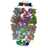
|
|---|---|
| 1 |
|
-Components
| #1: Protein | Mass: 56263.559 Da / Num. of mol.: 14 / Fragment: UNP residues 27-552 Source method: isolated from a genetically manipulated source Source: (gene. exp.) Homo sapiens (human) / Gene: HSPD1, HSP60 / Production host:  #2: Protein | Mass: 10744.400 Da / Num. of mol.: 14 Source method: isolated from a genetically manipulated source Source: (gene. exp.) Homo sapiens (human) / Gene: HSPE1 / Production host: #3: Chemical | ChemComp-ADP /   Mass: 427.201 Da / Num. of mol.: 14 / Source method: obtained synthetically / Formula: C10H15N5O10P2 / Comment: ADP, energy-carrying molecule*YM Mass: 427.201 Da / Num. of mol.: 14 / Source method: obtained synthetically / Formula: C10H15N5O10P2 / Comment: ADP, energy-carrying molecule*YM#4: Chemical | ChemComp-MG /   Mass: 24.305 Da / Num. of mol.: 14 / Source method: obtained synthetically / Formula: Mg Mass: 24.305 Da / Num. of mol.: 14 / Source method: obtained synthetically / Formula: Mg#5: Water | ChemComp-HOH / |  Mass: 18.015 Da / Num. of mol.: 98 / Source method: isolated from a natural source / Formula: H2O Mass: 18.015 Da / Num. of mol.: 98 / Source method: isolated from a natural source / Formula: H2OHas protein modification | N | |
|---|
-Experimental details
-Experiment
| Experiment | Method: ELECTRON MICROSCOPY |
|---|---|
| EM experiment | Aggregation state: PARTICLE / 3D reconstruction method: single particle reconstruction |
- Sample preparation
| Component | Name: Complex of Hsp60 with Hsp10 bound to ADP and Mg / Type: COMPLEX / Entity ID: #1-#2 / Source: RECOMBINANT |
|---|---|
| Molecular weight | Value: 0.98 MDa / Experimental value: NO |
| Source (natural) | Organism: Homo sapiens (human) |
| Source (recombinant) | Organism: |
| Buffer solution | pH: 7.7 |
| Specimen | Embedding applied: NO / Shadowing applied: NO / Staining applied: NO / Vitrification applied: YES |
| Specimen support | Grid material: GOLD / Grid type: Homemade |
| Vitrification | Instrument: HOMEMADE PLUNGER / Cryogen name: ETHANE |
- Electron microscopy imaging
| Experimental equipment |  Model: Titan Krios / Image courtesy: FEI Company |
|---|---|
| Microscopy | Model: FEI TITAN KRIOS |
| Electron gun | Electron source:  FIELD EMISSION GUN / Accelerating voltage: 300 kV / Illumination mode: FLOOD BEAM FIELD EMISSION GUN / Accelerating voltage: 300 kV / Illumination mode: FLOOD BEAM |
| Electron lens | Mode: BRIGHT FIELD / Alignment procedure: COMA FREE |
| Specimen holder | Cryogen: NITROGEN / Specimen holder model: FEI TITAN KRIOS AUTOGRID HOLDER |
| Image recording | Average exposure time: 10 sec. / Electron dose: 63 e/Å2 / Detector mode: COUNTING / Film or detector model: GATAN K2 SUMMIT (4k x 4k) |
- Processing
| Software | Name: PHENIX / Version: 1.13_2998: / Classification: refinement | ||||||||||||||||||||||||
|---|---|---|---|---|---|---|---|---|---|---|---|---|---|---|---|---|---|---|---|---|---|---|---|---|---|
| EM software |
| ||||||||||||||||||||||||
| CTF correction | Type: PHASE FLIPPING AND AMPLITUDE CORRECTION | ||||||||||||||||||||||||
| 3D reconstruction | Resolution: 3.08 Å / Resolution method: FSC 0.143 CUT-OFF / Num. of particles: 66013 / Symmetry type: POINT | ||||||||||||||||||||||||
| Refine LS restraints |
|
