 Movie
Movie Controller

[English] 日本語
 Yorodumi
- PDB-4qes: Structure of a 16 nm protein cage designed by fusing symmetric ol... -
+ Open data

- Basic information
| Entry | Database: PDB / ID: 4qes | ||||||
|---|---|---|---|---|---|---|---|
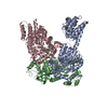
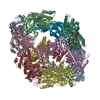
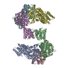
| Title | Structure of a 16 nm protein cage designed by fusing symmetric oligomeric domains, quadruple mutant, I222 form | ||||||
 Components Components | Non-haem bromoperoxidase BPO-A2, Matrix protein 1 chimera | ||||||
 Keywords Keywords | OXIDOREDUCTASE / VIRAL PROTEIN / protein design / bionanotechnology / protein assembly / symmetry / biomaterials | ||||||
| Function / homology |  Function and homology information Function and homology informationAssembly of Viral Components at the Budding Site / Influenza Infection / Fusion of the Influenza Virion to the Host Cell Endosome / Release / Budding / Packaging of Eight RNA Segments / Uncoating of the Influenza Virion / Entry of Influenza Virion into Host Cell via Endocytosis / Viral RNP Complexes in the Host Cell Nucleus / NEP/NS2 Interacts with the Cellular Export Machinery ...Assembly of Viral Components at the Budding Site / Influenza Infection / Fusion of the Influenza Virion to the Host Cell Endosome / Release / Budding / Packaging of Eight RNA Segments / Uncoating of the Influenza Virion / Entry of Influenza Virion into Host Cell via Endocytosis / Viral RNP Complexes in the Host Cell Nucleus / NEP/NS2 Interacts with the Cellular Export Machinery / Oxidoreductases; Acting on a peroxide as acceptor; Peroxidases / antibiotic biosynthetic process / Viral mRNA Translation / viral budding from plasma membrane / peroxidase activity / structural constituent of virion / host cell nucleus / virion membrane / RNA binding / extracellular region / plasma membrane Similarity search - Function | ||||||
| Biological species |  Streptomyces aureofaciens (bacteria) Streptomyces aureofaciens (bacteria)  Influenza A virus Influenza A virus | ||||||
| Method |  X-RAY DIFFRACTION / SYNCHROTRON / MOLECULAR REPLACEMENT / molecular replacement / Resolution: 4.193 Å X-RAY DIFFRACTION / SYNCHROTRON / MOLECULAR REPLACEMENT / molecular replacement / Resolution: 4.193 Å | ||||||
 Authors Authors | Lai, Y.-T. / Yeates, T.O. | ||||||
 Citation Citation | Journal: To be Published Title: Structural transition of a protein nanocage as a function of pH and salt concentration. Authors: Lai, Y.-T. / Yeates, T.O. | ||||||
| History |
|
- Structure visualization
| Structure viewer | Molecule: MolmilJmol/JSmol |
|---|
- Downloads & links
-Download
| PDBx/mmCIF format | 4qes.cif.gz | 256.3 KB | Display | PDBx/mmCIF format |
|---|---|---|---|---|
| PDB format | pdb4qes.ent.gz | 209.3 KB | Display | PDB format |
| PDBx/mmJSON format | 4qes.json.gz | Tree view | PDBx/mmJSON format | |
| Others |  Other downloads Other downloads |
-Validation report
| Arichive directory | https://data.pdbj.org/pub/pdb/validation_reports/qe/4qesftp://data.pdbj.org/pub/pdb/validation_reports/qe/4qes | HTTPS FTP |
|---|
-Related structure data


-Links
 PDBj
PDBj


- Assembly
| Deposited unit | 
| |||||||||||||||||||||||||||||||||||||||||||||||||||||
|---|---|---|---|---|---|---|---|---|---|---|---|---|---|---|---|---|---|---|---|---|---|---|---|---|---|---|---|---|---|---|---|---|---|---|---|---|---|---|---|---|---|---|---|---|---|---|---|---|---|---|---|---|---|---|
| 1 | 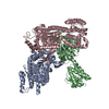
| |||||||||||||||||||||||||||||||||||||||||||||||||||||
| Unit cell |
| |||||||||||||||||||||||||||||||||||||||||||||||||||||
| Noncrystallographic symmetry (NCS) | NCS domain:
NCS domain segments: Component-ID: _ / Beg auth comp-ID: PRO / Beg label comp-ID: PRO / End auth comp-ID: GLN / End label comp-ID: GLN / Refine code: _ / Auth seq-ID: 1 - 441 / Label seq-ID: 2 - 442
NCS ensembles :
|
-Components
| #1: Protein | Mass: 50189.266 Da / Num. of mol.: 3 / Fragment: SEE REMARK 999 / Mutation: K118A, L279Q, Q24T, Y51A Source method: isolated from a genetically manipulated source Source: (gene. exp.) Streptomyces aureofaciens (bacteria), (gene. exp.) Influenza A virusGene: bpoA2, M / Plasmid: pET22b / Production host: References: UniProt: P29715, UniProt: P03485, Oxidoreductases; Acting on a peroxide as acceptor; Peroxidases Sequence details | THE DESIGNED PROTEIN CONSTRUCT IS A CHIMERA COMPRISING BPO2 (UNP P29715) AND RESIDUES 2-164 (UNP ...THE DESIGNED PROTEIN CONSTRUCT IS A CHIMERA COMPRISING | |
|---|
-Experimental details
-Experiment
| Experiment | Method: X-RAY DIFFRACTION / Number of used crystals: 1 |
|---|
- Sample preparation
| Crystal | Density Matthews: 2.85 Å3/Da / Density % sol: 56.78 % |
|---|---|
| Crystal grow | Temperature: 298 K / Method: vapor diffusion, hanging drop / pH: 4.4 Details: 0.1 M sodium citrate, pH 4.4, 11% PEG3000, 200 mM sodium chloride, VAPOR DIFFUSION, HANGING DROP, temperature 298K |
-Data collection
| Diffraction | Mean temperature: 100 K | |||||||||||||||||||||||||||||||||||||||||||||||||||||||||||||||||||||||||||||||||||||||||||||||||||||||||||||||||||||||||||||||||||||||||||||||||||
|---|---|---|---|---|---|---|---|---|---|---|---|---|---|---|---|---|---|---|---|---|---|---|---|---|---|---|---|---|---|---|---|---|---|---|---|---|---|---|---|---|---|---|---|---|---|---|---|---|---|---|---|---|---|---|---|---|---|---|---|---|---|---|---|---|---|---|---|---|---|---|---|---|---|---|---|---|---|---|---|---|---|---|---|---|---|---|---|---|---|---|---|---|---|---|---|---|---|---|---|---|---|---|---|---|---|---|---|---|---|---|---|---|---|---|---|---|---|---|---|---|---|---|---|---|---|---|---|---|---|---|---|---|---|---|---|---|---|---|---|---|---|---|---|---|---|---|---|---|
| Diffraction source | Source: SYNCHROTRON / Site: APS  / Beamline: 24-ID-C / Wavelength: 0.9789 Å / Beamline: 24-ID-C / Wavelength: 0.9789 Å | |||||||||||||||||||||||||||||||||||||||||||||||||||||||||||||||||||||||||||||||||||||||||||||||||||||||||||||||||||||||||||||||||||||||||||||||||||
| Detector | Type: DECTRIS PILATUS 6M-F / Detector: PIXEL / Date: Feb 28, 2014 | |||||||||||||||||||||||||||||||||||||||||||||||||||||||||||||||||||||||||||||||||||||||||||||||||||||||||||||||||||||||||||||||||||||||||||||||||||
| Radiation | Monochromator: Cryo-Cooled double crystal Si(111) / Protocol: SINGLE WAVELENGTH / Monochromatic (M) / Laue (L): M / Scattering type: x-ray | |||||||||||||||||||||||||||||||||||||||||||||||||||||||||||||||||||||||||||||||||||||||||||||||||||||||||||||||||||||||||||||||||||||||||||||||||||
| Radiation wavelength | Wavelength: 0.9789 Å / Relative weight: 1 | |||||||||||||||||||||||||||||||||||||||||||||||||||||||||||||||||||||||||||||||||||||||||||||||||||||||||||||||||||||||||||||||||||||||||||||||||||
| Reflection | Resolution: 4.19→83.835 Å / Num. obs: 12914 / % possible obs: 99.7 % / Observed criterion σ(I): -3 / Biso Wilson estimate: 161.223 Å2 / Rmerge(I) obs: 0.127 / Χ2: 0.824 / Net I/σ(I): 14.16 | |||||||||||||||||||||||||||||||||||||||||||||||||||||||||||||||||||||||||||||||||||||||||||||||||||||||||||||||||||||||||||||||||||||||||||||||||||
| Reflection shell |
|
-Phasing
| Phasing | Method: molecular replacement | |||||||||
|---|---|---|---|---|---|---|---|---|---|---|
| Phasing MR | Model details: Phaser MODE: MR_AUTO
|
- Processing
| Software |
| |||||||||||||||||||||||||||||||||||||||||||||||||||||||||||||||||||||||||||
|---|---|---|---|---|---|---|---|---|---|---|---|---|---|---|---|---|---|---|---|---|---|---|---|---|---|---|---|---|---|---|---|---|---|---|---|---|---|---|---|---|---|---|---|---|---|---|---|---|---|---|---|---|---|---|---|---|---|---|---|---|---|---|---|---|---|---|---|---|---|---|---|---|---|---|---|---|
| Refinement | Method to determine structure: MOLECULAR REPLACEMENT / Resolution: 4.193→83.83 Å / Cor.coef. Fo:Fc: 0.911 / Cor.coef. Fo:Fc free: 0.938 / WRfactor Rfree: 0.2867 / WRfactor Rwork: 0.2438 / FOM work R set: 0.737 / SU B: 76.252 / SU ML: 0.981 / SU Rfree: 1.1442 / Cross valid method: THROUGHOUT / σ(F): 0 / ESU R Free: 1.144 / Stereochemistry target values: MAXIMUM LIKELIHOOD / Details: HYDROGENS HAVE BEEN ADDED IN THE RIDING POSITIONS
| |||||||||||||||||||||||||||||||||||||||||||||||||||||||||||||||||||||||||||
| Solvent computation | Ion probe radii: 0.8 Å / Shrinkage radii: 0.8 Å / VDW probe radii: 1.2 Å / Solvent model: MASK | |||||||||||||||||||||||||||||||||||||||||||||||||||||||||||||||||||||||||||
| Displacement parameters | Biso max: 500 Å2 / Biso mean: 198.022 Å2 / Biso min: 87.77 Å2
| |||||||||||||||||||||||||||||||||||||||||||||||||||||||||||||||||||||||||||
| Refinement step | Cycle: LAST / Resolution: 4.193→83.83 Å
| |||||||||||||||||||||||||||||||||||||||||||||||||||||||||||||||||||||||||||
| Refine LS restraints |
| |||||||||||||||||||||||||||||||||||||||||||||||||||||||||||||||||||||||||||
| Refine LS restraints NCS | Refine-ID: X-RAY DIFFRACTION / Type: interatomic distance / Weight position: 0.05
| |||||||||||||||||||||||||||||||||||||||||||||||||||||||||||||||||||||||||||
| LS refinement shell | Resolution: 4.193→4.302 Å / Total num. of bins used: 20
|
