 Movie
Movie Controller

[English] 日本語
 Yorodumi
- PDB-8eog: Structure of the human L-type voltage-gated calcium channel Cav1.... -
+ Open data

- Basic information
| Entry | Database: PDB / ID: 8eog | |||||||||
|---|---|---|---|---|---|---|---|---|---|---|
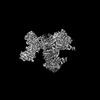
| Title | Structure of the human L-type voltage-gated calcium channel Cav1.2 complexed with L-leucine | |||||||||
 Components Components |
| |||||||||
 Keywords Keywords | MEMBRANE PROTEIN / voltage-gated calcium channel / CaV alpha2delta / drug binding / gabapentinoid | |||||||||
| Function / homology |  Function and homology information Function and homology informationpositive regulation of high voltage-gated calcium channel activity / calcium ion transmembrane transport via high voltage-gated calcium channel / L-type voltage-gated calcium channel complex / calcium ion import across plasma membrane / voltage-gated calcium channel activity / T-tubule / calcium channel regulator activity / metal ion binding / cytoplasm Similarity search - Function | |||||||||
| Biological species |   Homo sapiens (human) Homo sapiens (human) | |||||||||
| Method | ELECTRON MICROSCOPY / single particle reconstruction / cryo EM / Resolution: 3.3 Å | |||||||||
 Authors Authors | Chen, Z. / Mondal, A. / Abderemane-Ali, F. / Minor, D.L. | |||||||||
| Funding support |  United States, 2items United States, 2items
| |||||||||
 Citation Citation | Journal: Nature / Year: 2023 Title: EMC chaperone-Ca structure reveals an ion channel assembly intermediate. Authors: Zhou Chen / Abhisek Mondal / Fayal Abderemane-Ali / Seil Jang / Sangeeta Niranjan / José L Montaño / Balyn W Zaro / Daniel L Minor / Abstract: Voltage-gated ion channels (VGICs) comprise multiple structural units, the assembly of which is required for function. Structural understanding of how VGIC subunits assemble and whether chaperone ...Voltage-gated ion channels (VGICs) comprise multiple structural units, the assembly of which is required for function. Structural understanding of how VGIC subunits assemble and whether chaperone proteins are required is lacking. High-voltage-activated calcium channels (Cas) are paradigmatic multisubunit VGICs whose function and trafficking are powerfully shaped by interactions between pore-forming Ca1 or Ca2 Caα (ref. ), and the auxiliary Caβ and Caαδ subunits. Here we present cryo-electron microscopy structures of human brain and cardiac Ca1.2 bound with Caβ to a chaperone-the endoplasmic reticulum membrane protein complex (EMC)-and of the assembled Ca1.2-Caβ-Caαδ-1 channel. These structures provide a view of an EMC-client complex and define EMC sites-the transmembrane (TM) and cytoplasmic (Cyto) docks; interaction between these sites and the client channel causes partial extraction of a pore subunit and splays open the Caαδ-interaction site. The structures identify the Caαδ-binding site for gabapentinoid anti-pain and anti-anxiety drugs, show that EMC and Caαδ interactions with the channel are mutually exclusive, and indicate that EMC-to-Caαδ hand-off involves a divalent ion-dependent step and Ca1.2 element ordering. Disruption of the EMC-Ca complex compromises Ca function, suggesting that the EMC functions as a channel holdase that facilitates channel assembly. Together, the structures reveal a Ca assembly intermediate and EMC client-binding sites that could have wide-ranging implications for the biogenesis of VGICs and other membrane proteins. | |||||||||
| History |
|
- Structure visualization
| Structure viewer | Molecule: MolmilJmol/JSmol |
|---|
- Downloads & links
-Download
| PDBx/mmCIF format | 8eog.cif.gz | 462.6 KB | Display | PDBx/mmCIF format |
|---|---|---|---|---|
| PDB format | pdb8eog.ent.gz | 361 KB | Display | PDB format |
| PDBx/mmJSON format | 8eog.json.gz | Tree view | PDBx/mmJSON format | |
| Others |  Other downloads Other downloads |
-Validation report
| Arichive directory | https://data.pdbj.org/pub/pdb/validation_reports/eo/8eogftp://data.pdbj.org/pub/pdb/validation_reports/eo/8eog | HTTPS FTP |
|---|
-Related structure data
| Related structure data | 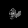 28375MC 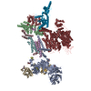 8eoiC M: map data used to model this data C: citing same article ( |
|---|---|
| Similar structure data |


-Links
 PDBj
PDBj


- Assembly
| Deposited unit | 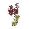
|
|---|---|
| 1 |
|
-Components
-Voltage-dependent ... , 2 types, 2 molecules DC
| #1: Protein | Mass: 118941.695 Da / Num. of mol.: 1 Source method: isolated from a genetically manipulated source Source: (gene. exp.) Homo sapiens (human) / References: UniProt: P13806 |
|---|---|
| #3: Protein | Mass: 21667.154 Da / Num. of mol.: 1 Source method: isolated from a genetically manipulated source Source: (gene. exp.) Homo sapiens (human) / References: UniProt: P54286 |
-Protein , 1 types, 1 molecules K
| #2: Protein | Mass: 170774.938 Da / Num. of mol.: 1 Source method: isolated from a genetically manipulated source Source: (gene. exp.) Homo sapiens (human) / Gene: CACNA1C, CACH2, CACN2, CACNL1A1, CCHL1A1 / Production host: Homo sapiens (human) / References: UniProt: Q13936-20 |
|---|
-Sugars , 3 types, 8 molecules 
| #4: Polysaccharide | 2-acetamido-2-deoxy-beta-D-glucopyranose-(1-4)-2-acetamido-2-deoxy-beta-D-glucopyranose-(1-4)-2- ...2-acetamido-2-deoxy-beta-D-glucopyranose-(1-4)-2-acetamido-2-deoxy-beta-D-glucopyranose-(1-4)-2-acetamido-2-deoxy-beta-D-glucopyranose Source method: isolated from a genetically manipulated source | ||
|---|---|---|---|
| #5: Polysaccharide | Source method: isolated from a genetically manipulated source #6: Sugar | ChemComp-NAG /  Type: D-saccharide, beta linking / Mass: 221.208 Da / Num. of mol.: 5 / Source method: obtained synthetically / Formula: C8H15NO6 Type: D-saccharide, beta linking / Mass: 221.208 Da / Num. of mol.: 5 / Source method: obtained synthetically / Formula: C8H15NO6 |
-Non-polymers , 7 types, 13 molecules 


| #7: Chemical |  Mass: 40.078 Da / Num. of mol.: 3 / Source method: obtained synthetically / Formula: Ca Mass: 40.078 Da / Num. of mol.: 3 / Source method: obtained synthetically / Formula: Ca#8: Chemical | ChemComp-NA / |  Mass: 22.990 Da / Num. of mol.: 1 / Source method: isolated from a natural source / Formula: Na Mass: 22.990 Da / Num. of mol.: 1 / Source method: isolated from a natural source / Formula: Na#9: Chemical | ChemComp-LEU / |  Type: L-peptide linking / Mass: 131.173 Da / Num. of mol.: 1 / Source method: obtained synthetically / Formula: C6H13NO2 Type: L-peptide linking / Mass: 131.173 Da / Num. of mol.: 1 / Source method: obtained synthetically / Formula: C6H13NO2#10: Chemical |  Mass: 386.654 Da / Num. of mol.: 3 / Source method: obtained synthetically / Formula: C27H46O Mass: 386.654 Da / Num. of mol.: 3 / Source method: obtained synthetically / Formula: C27H46O#11: Chemical | ChemComp-WNZ / ( |  Mass: 579.746 Da / Num. of mol.: 1 / Source method: obtained synthetically / Formula: C29H58NO8P Mass: 579.746 Da / Num. of mol.: 1 / Source method: obtained synthetically / Formula: C29H58NO8P#12: Chemical | ChemComp-WO9 / ( |  Mass: 649.879 Da / Num. of mol.: 1 / Source method: obtained synthetically / Formula: C34H68NO8P Mass: 649.879 Da / Num. of mol.: 1 / Source method: obtained synthetically / Formula: C34H68NO8P#13: Water | ChemComp-HOH / | Mass: 18.015 Da / Num. of mol.: 3 / Source method: isolated from a natural source / Formula: H2O |
|---|
-Details
| Has ligand of interest | N |
|---|---|
| Has protein modification | Y |
-Experimental details
-Experiment
| Experiment | Method: ELECTRON MICROSCOPY |
|---|---|
| EM experiment | Aggregation state: PARTICLE / 3D reconstruction method: single particle reconstruction |
- Sample preparation
| Component |
| ||||||||||||||||||||||||||||||
|---|---|---|---|---|---|---|---|---|---|---|---|---|---|---|---|---|---|---|---|---|---|---|---|---|---|---|---|---|---|---|---|
| Molecular weight |
| ||||||||||||||||||||||||||||||
| Source (natural) |
| ||||||||||||||||||||||||||||||
| Source (recombinant) |
| ||||||||||||||||||||||||||||||
| Buffer solution | pH: 8 | ||||||||||||||||||||||||||||||
| Specimen | Conc.: 2.7 mg/ml / Embedding applied: NO / Shadowing applied: NO / Staining applied: NO / Vitrification applied: YES | ||||||||||||||||||||||||||||||
| Specimen support | Grid material: GOLD / Grid mesh size: 300 divisions/in. / Grid type: Quantifoil R1.2/1.3 | ||||||||||||||||||||||||||||||
| Vitrification | Instrument: FEI VITROBOT MARK IV / Cryogen name: ETHANE / Humidity: 100 % / Chamber temperature: 277 K |
- Electron microscopy imaging
| Experimental equipment |  Model: Titan Krios / Image courtesy: FEI Company |
|---|---|
| Microscopy | Model: FEI TITAN KRIOS |
| Electron gun | Electron source:  FIELD EMISSION GUN / Accelerating voltage: 300 kV / Illumination mode: FLOOD BEAM FIELD EMISSION GUN / Accelerating voltage: 300 kV / Illumination mode: FLOOD BEAM |
| Electron lens | Mode: BRIGHT FIELD / Nominal magnification: 105000 X / Nominal defocus max: 1700 nm / Nominal defocus min: 900 nm / Cs: 2.7 mm |
| Image recording | Electron dose: 46 e/Å2 / Film or detector model: GATAN K3 (6k x 4k) |
- Processing
| Software | Name: PHENIX / Version: 1.20.1_4487: / Classification: refinement | ||||||||||||||||||||||||
|---|---|---|---|---|---|---|---|---|---|---|---|---|---|---|---|---|---|---|---|---|---|---|---|---|---|
| CTF correction | Type: PHASE FLIPPING AND AMPLITUDE CORRECTION | ||||||||||||||||||||||||
| Symmetry | Point symmetry: C1 (asymmetric) | ||||||||||||||||||||||||
| 3D reconstruction | Resolution: 3.3 Å / Resolution method: FSC 0.143 CUT-OFF / Num. of particles: 269802 / Symmetry type: POINT | ||||||||||||||||||||||||
| Refine LS restraints |
|
