Movie
Movie Controller

[English] 日本語
 Yorodumi
- EMDB-28376: Structure of a human EMC:human Cav1.2 channel complex in GDN dete... -
+ Open data

- Basic information
| Entry |  | |||||||||
|---|---|---|---|---|---|---|---|---|---|---|
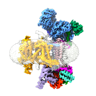
| Title | Structure of a human EMC:human Cav1.2 channel complex in GDN detergent (ECAB Map 3) | |||||||||
 Map data Map data | Structure of a human EMC:human Cav1.2 channel complex in GDN detergent (ECAB Map 3) | |||||||||
 Sample Sample |
| |||||||||
 Keywords Keywords | endoplasmic reticulum membrane protein complex / voltage-gated calcium channel / holdase / biogenesis / MEMBRANE PROTEIN | |||||||||
| Function / homology |  Function and homology information Function and homology informationextrinsic component of endoplasmic reticulum membrane / : / EMC complex / positive regulation of high voltage-gated calcium channel activity / omegasome membrane / voltage-gated calcium channel activity involved in AV node cell action potential / voltage-gated calcium channel activity involved in cardiac muscle cell action potential / protein insertion into ER membrane by stop-transfer membrane-anchor sequence / immune system development / positive regulation of adenylate cyclase activity ...extrinsic component of endoplasmic reticulum membrane / : / EMC complex / positive regulation of high voltage-gated calcium channel activity / omegasome membrane / voltage-gated calcium channel activity involved in AV node cell action potential / voltage-gated calcium channel activity involved in cardiac muscle cell action potential / protein insertion into ER membrane by stop-transfer membrane-anchor sequence / immune system development / positive regulation of adenylate cyclase activity / membrane depolarization during atrial cardiac muscle cell action potential / magnesium ion transport / calcium ion transmembrane transport via high voltage-gated calcium channel / tail-anchored membrane protein insertion into ER membrane / Miscellaneous transport and binding events / cobalt ion transmembrane transporter activity / Phase 2 - plateau phase / ferrous iron transmembrane transporter activity / high voltage-gated calcium channel activity / copper ion transport / magnesium ion transmembrane transporter activity / cardiac conduction / embryonic forelimb morphogenesis / membrane depolarization during AV node cell action potential / L-type voltage-gated calcium channel complex / membrane depolarization during cardiac muscle cell action potential / cell communication by electrical coupling involved in cardiac conduction / positive regulation of muscle contraction / NCAM1 interactions / camera-type eye development / regulation of ventricular cardiac muscle cell action potential / cardiac muscle cell action potential involved in contraction / calcium ion transport into cytosol / voltage-gated calcium channel complex / Phase 0 - rapid depolarisation / regulation of heart rate by cardiac conduction / alpha-actinin binding / calcium ion import across plasma membrane / RHOA GTPase cycle / autophagosome assembly / voltage-gated calcium channel activity / regulation of cardiac muscle contraction by regulation of the release of sequestered calcium ion / positive regulation of endothelial cell proliferation / positive regulation of endothelial cell migration / calcium channel regulator activity / Regulation of insulin secretion / postsynaptic density membrane / Z disc / calcium ion transmembrane transport / positive regulation of angiogenesis / Adrenaline,noradrenaline inhibits insulin secretion / heart development / positive regulation of cytosolic calcium ion concentration / carbohydrate binding / angiogenesis / early endosome membrane / perikaryon / early endosome / calmodulin binding / postsynaptic density / Golgi membrane / apoptotic process / dendrite / endoplasmic reticulum membrane / Golgi apparatus / endoplasmic reticulum / protein-containing complex / extracellular region / membrane / metal ion binding / plasma membrane / cytoplasm Similarity search - Function | |||||||||
| Biological species |  Homo sapiens (human) / Homo sapiens (human) /  | |||||||||
| Method | single particle reconstruction / cryo EM / Resolution: 3.4 Å | |||||||||
 Authors Authors | Chen Z / Mondal A / Abderemane-Ali F / Minor DL | |||||||||
| Funding support |  United States, 2 items United States, 2 items
| |||||||||
 Citation Citation | Journal: Nature / Year: 2023 Title: EMC chaperone-Ca structure reveals an ion channel assembly intermediate. Authors: Zhou Chen / Abhisek Mondal / Fayal Abderemane-Ali / Seil Jang / Sangeeta Niranjan / José L Montaño / Balyn W Zaro / Daniel L Minor / Abstract: Voltage-gated ion channels (VGICs) comprise multiple structural units, the assembly of which is required for function. Structural understanding of how VGIC subunits assemble and whether chaperone ...Voltage-gated ion channels (VGICs) comprise multiple structural units, the assembly of which is required for function. Structural understanding of how VGIC subunits assemble and whether chaperone proteins are required is lacking. High-voltage-activated calcium channels (Cas) are paradigmatic multisubunit VGICs whose function and trafficking are powerfully shaped by interactions between pore-forming Ca1 or Ca2 Caα (ref. ), and the auxiliary Caβ and Caαδ subunits. Here we present cryo-electron microscopy structures of human brain and cardiac Ca1.2 bound with Caβ to a chaperone-the endoplasmic reticulum membrane protein complex (EMC)-and of the assembled Ca1.2-Caβ-Caαδ-1 channel. These structures provide a view of an EMC-client complex and define EMC sites-the transmembrane (TM) and cytoplasmic (Cyto) docks; interaction between these sites and the client channel causes partial extraction of a pore subunit and splays open the Caαδ-interaction site. The structures identify the Caαδ-binding site for gabapentinoid anti-pain and anti-anxiety drugs, show that EMC and Caαδ interactions with the channel are mutually exclusive, and indicate that EMC-to-Caαδ hand-off involves a divalent ion-dependent step and Ca1.2 element ordering. Disruption of the EMC-Ca complex compromises Ca function, suggesting that the EMC functions as a channel holdase that facilitates channel assembly. Together, the structures reveal a Ca assembly intermediate and EMC client-binding sites that could have wide-ranging implications for the biogenesis of VGICs and other membrane proteins. | |||||||||
| History |
|
- Structure visualization
| Supplemental images |
|---|
- Downloads & links
-EMDB archive
| Map data | emd_28376.map.gz | 260.4 MB | EMDB map data format | |
|---|---|---|---|---|
| Header (meta data) | emd-28376-v30.xmlemd-28376.xml | 28.1 KB 28.1 KB | Display Display | EMDB header |
| Images |  emd_28376.png emd_28376.png | 132 KB | ||
| Filedesc metadata | emd-28376.cif.gz | 9.4 KB | ||
| Archive directory |  http://ftp.pdbj.org/pub/emdb/structures/EMD-28376ftp://ftp.pdbj.org/pub/emdb/structures/EMD-28376 http://ftp.pdbj.org/pub/emdb/structures/EMD-28376ftp://ftp.pdbj.org/pub/emdb/structures/EMD-28376 | HTTPS FTP |
-Related structure data
| Related structure data |  8eoiMC  8eogC M: atomic model generated by this map C: citing same article ( |
|---|---|
| Similar structure data |


-Links
| EMDB pages | EMDB (EBI/PDBe) / EMDataResource |
|---|---|
| Related items in Molecule of the Month |


-Map
| File | Download / File: emd_28376.map.gz / Format: CCP4 / Size: 325 MB / Type: IMAGE STORED AS FLOATING POINT NUMBER (4 BYTES) | ||||||||||||||||||||||||||||||||||||
|---|---|---|---|---|---|---|---|---|---|---|---|---|---|---|---|---|---|---|---|---|---|---|---|---|---|---|---|---|---|---|---|---|---|---|---|---|---|
| Annotation | Structure of a human EMC:human Cav1.2 channel complex in GDN detergent (ECAB Map 3) | ||||||||||||||||||||||||||||||||||||
| Projections & slices | Image control
Images are generated by Spider. | ||||||||||||||||||||||||||||||||||||
| Voxel size | X=Y=Z: 0.8466 Å | ||||||||||||||||||||||||||||||||||||
| Density |
| ||||||||||||||||||||||||||||||||||||
| Symmetry | Space group: 1 | ||||||||||||||||||||||||||||||||||||
| Details | EMDB XML:
|
 X (Sec.)
 Y (Row.)
 Z (Col.)


-Supplemental data
- Sample components
+Entire : Ternary complex of the human ER membrane protein complex (EMC) wi...
+Supramolecule #1: Ternary complex of the human ER membrane protein complex (EMC) wi...
+Supramolecule #2: Human CaV alpha1C
+Supramolecule #3: ER membrane protein complex subunit 1
+Supramolecule #4: Rabbit CaV beta3
+Macromolecule #1: ER membrane protein complex subunit 1
+Macromolecule #2: ER membrane protein complex subunit 2
+Macromolecule #3: ER membrane protein complex subunit 3
+Macromolecule #4: ER membrane protein complex subunit 5
+Macromolecule #5: ER membrane protein complex subunit 6
+Macromolecule #6: ER membrane protein complex subunit 7
+Macromolecule #7: ER membrane protein complex subunit 8
+Macromolecule #8: ER membrane protein complex subunit 10
+Macromolecule #9: Voltage-dependent L-type calcium channel subunit alpha-1C
+Macromolecule #10: Voltage-dependent L-type calcium channel subunit beta-3
+Macromolecule #11: ER membrane protein complex subunit 4
+Macromolecule #13: 2-acetamido-2-deoxy-beta-D-glucopyranose

+Macromolecule #14: (3beta,14beta,17beta,25R)-3-[4-methoxy-3-(methoxymethyl)butoxy]sp...
-Experimental details
-Structure determination
| Method | cryo EM |
|---|---|
 Processing Processing | single particle reconstruction |
| Aggregation state | particle |
-Sample preparation
| Concentration | 1.7 mg/mL |
|---|---|
| Buffer | pH: 8 |
| Grid | Model: Quantifoil R1.2/1.3 / Material: GOLD / Mesh: 300 / Support film - Material: CARBON / Support film - topology: HOLEY |
| Vitrification | Cryogen name: ETHANE / Chamber humidity: 100 % / Chamber temperature: 277 K / Instrument: FEI VITROBOT MARK IV |
- Electron microscopy
| Microscope | FEI TITAN KRIOS |
|---|---|
| Image recording | Film or detector model: GATAN K3 (6k x 4k) / Average electron dose: 46.0 e/Å2 |
| Electron beam | Acceleration voltage: 300 kV / Electron source:  FIELD EMISSION GUN FIELD EMISSION GUN |
| Electron optics | Illumination mode: FLOOD BEAM / Imaging mode: BRIGHT FIELD / Cs: 2.7 mm / Nominal defocus max: 1.7 µm / Nominal defocus min: 0.9 µm / Nominal magnification: 105000 |
| Experimental equipment |  Model: Titan Krios / Image courtesy: FEI Company |
-Image processing
| Startup model | Type of model: PDB ENTRY PDB model - PDB ID: Details: 6WW7 (PDB ID) and 7MIY (PDB ID) used to generate the startup model. |
|---|---|
| Final reconstruction | Applied symmetry - Point group: C1 (asymmetric) / Resolution.type: BY AUTHOR / Resolution: 3.4 Å / Resolution method: FSC 0.143 CUT-OFF / Number images used: 487067 |
| Initial angle assignment | Type: MAXIMUM LIKELIHOOD |
| Final angle assignment | Type: MAXIMUM LIKELIHOOD |