National Institutes of Health/National Heart, Lung, and Blood Institute (NIH/NHLBI)
HL080050
United States
National Institutes of Health/National Institute on Deafness and Other Communication Disorders (NIH/NIDCD)
DC007664
United States
Citation
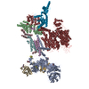
Journal: Nature / Year: 2023 Title: EMC chaperone-Ca structure reveals an ion channel assembly intermediate. Authors: Zhou Chen / Abhisek Mondal / Fayal Abderemane-Ali / Seil Jang / Sangeeta Niranjan / José L Montaño / Balyn W Zaro / Daniel L Minor / Abstract: Voltage-gated ion channels (VGICs) comprise multiple structural units, the assembly of which is required for function. Structural understanding of how VGIC subunits assemble and whether chaperone ...Voltage-gated ion channels (VGICs) comprise multiple structural units, the assembly of which is required for function. Structural understanding of how VGIC subunits assemble and whether chaperone proteins are required is lacking. High-voltage-activated calcium channels (Cas) are paradigmatic multisubunit VGICs whose function and trafficking are powerfully shaped by interactions between pore-forming Ca1 or Ca2 Caα (ref. ), and the auxiliary Caβ and Caαδ subunits. Here we present cryo-electron microscopy structures of human brain and cardiac Ca1.2 bound with Caβ to a chaperone-the endoplasmic reticulum membrane protein complex (EMC)-and of the assembled Ca1.2-Caβ-Caαδ-1 channel. These structures provide a view of an EMC-client complex and define EMC sites-the transmembrane (TM) and cytoplasmic (Cyto) docks; interaction between these sites and the client channel causes partial extraction of a pore subunit and splays open the Caαδ-interaction site. The structures identify the Caαδ-binding site for gabapentinoid anti-pain and anti-anxiety drugs, show that EMC and Caαδ interactions with the channel are mutually exclusive, and indicate that EMC-to-Caαδ hand-off involves a divalent ion-dependent step and Ca1.2 element ordering. Disruption of the EMC-Ca complex compromises Ca function, suggesting that the EMC functions as a channel holdase that facilitates channel assembly. Together, the structures reveal a Ca assembly intermediate and EMC client-binding sites that could have wide-ranging implications for the biogenesis of VGICs and other membrane proteins.
In the structure databanks used in Yorodumi, some data are registered as the other names, "COVID-19 virus" and "2019-nCoV". Here are the details of the virus and the list of structure data.
Jan 31, 2019. EMDB accession codes are about to change! (news from PDBe EMDB page)
EMDB accession codes are about to change! (news from PDBe EMDB page)
The allocation of 4 digits for EMDB accession codes will soon come to an end. Whilst these codes will remain in use, new EMDB accession codes will include an additional digit and will expand incrementally as the available range of codes is exhausted. The current 4-digit format prefixed with “EMD-” (i.e. EMD-XXXX) will advance to a 5-digit format (i.e. EMD-XXXXX), and so on. It is currently estimated that the 4-digit codes will be depleted around Spring 2019, at which point the 5-digit format will come into force.
The EM Navigator/Yorodumi systems omit the EMD- prefix.
Related info.:Q: What is EMD? / ID/Accession-code notation in Yorodumi/EM Navigator
Yorodumi is a browser for structure data from EMDB, PDB, SASBDB, etc.
This page is also the successor to EM Navigator detail page, and also detail information page/front-end page for Omokage search.
The word "yorodu" (or yorozu) is an old Japanese word meaning "ten thousand". "mi" (miru) is to see.
Related info.:EMDB / PDB / SASBDB / Comparison of 3 databanks / Yorodumi Search / Aug 31, 2016. New EM Navigator & Yorodumi / Yorodumi Papers / Jmol/JSmol / Function and homology information / Changes in new EM Navigator and Yorodumi
 Movie
Movie Controller

 Yorodumi
 Open data
 Basic information
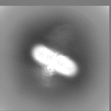
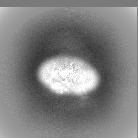
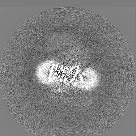
 Map data
 Sample
 Keywords
 Homo sapiens (human) /
 Authors
 United States, 2 items
  Citation
 Structure visualization
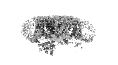
 Downloads & links
 EMDB map data format
 emd_28564.png
 http://ftp.pdbj.org/pub/emdb/structures/EMD-28564


 Z (Sec.)
 Y (Row.)
 X (Col.)


 Sample components
 Processing
 Electron microscopy
 FIELD EMISSION GUN

