 Movie
Movie Controller


+ Open data

- Basic information
| Entry | Database: PDB / ID: 8bqa | |||||||||
|---|---|---|---|---|---|---|---|---|---|---|
| Title | CjCel5B endo-glucanase bound to CB665 covalent inhibitor | |||||||||
 Components Components | Endoglucanase | |||||||||
 Keywords Keywords | HYDROLASE / Carbohydrate / inhibitor / covalent / cellulase | |||||||||
| Function / homology |  Function and homology information Function and homology informationcellulose binding / cellulase / cellulase activity / cellulose catabolic process Similarity search - Function | |||||||||
| Biological species |  Cellvibrio japonicus (bacteria) Cellvibrio japonicus (bacteria) | |||||||||
| Method |  X-RAY DIFFRACTION / SYNCHROTRON / MOLECULAR REPLACEMENT / Resolution: 1.67 Å X-RAY DIFFRACTION / SYNCHROTRON / MOLECULAR REPLACEMENT / Resolution: 1.67 Å | |||||||||
 Authors Authors | McGregor, N.G.S. / de Boer, C. / Overkleeft, H.S. / Davies, G.J. | |||||||||
| Funding support |  United Kingdom, European Union, 2items United Kingdom, European Union, 2items
| |||||||||
 Citation Citation | Journal: Acs Cent.Sci. / Year: 2023 Title: A Multiplexing Activity-Based Protein-Profiling Platform for Dissection of a Native Bacterial Xyloglucan-Degrading System. Authors: McGregor, N.G.S. / de Boer, C. / Foucart, Q.P.O. / Beenakker, T. / Offen, W.A. / Codee, J.D.C. / Willems, L.I. / Overkleeft, H.S. / Davies, G.J. | |||||||||
| History |
|
- Structure visualization
| Structure viewer | Molecule: MolmilJmol/JSmol |
|---|
- Downloads & links
-Download
| PDBx/mmCIF format | 8bqa.cif.gz | 130.7 KB | Display | PDBx/mmCIF format |
|---|---|---|---|---|
| PDB format | pdb8bqa.ent.gz | Display | PDB format | |
| PDBx/mmJSON format | 8bqa.json.gz | Tree view | PDBx/mmJSON format | |
| Others |  Other downloads Other downloads |
-Validation report
| Arichive directory | https://data.pdbj.org/pub/pdb/validation_reports/bq/8bqaftp://data.pdbj.org/pub/pdb/validation_reports/bq/8bqa | HTTPS FTP |
|---|
-Related structure data
| Related structure data |  8bn7C 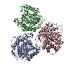 8bqbC  8bqcC  8oz1C 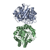 1tvnS S: Starting model for refinement C: citing same article ( |
|---|---|
| Similar structure data |
-Links
 PDBj
PDBj
- Assembly
| Deposited unit | 
| ||||||||
|---|---|---|---|---|---|---|---|---|---|
| 1 |
| ||||||||
| Unit cell |
|
-Components
| #1: Protein | Mass: 33519.051 Da / Num. of mol.: 1 Source method: isolated from a genetically manipulated source Source: (gene. exp.) Cellvibrio japonicus (bacteria) / Gene: cel5B, CJA_2983 / Production host: |
|---|---|
| #2: Chemical | ChemComp-RBH / ~{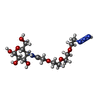  Mass: 395.386 Da / Num. of mol.: 1 / Source method: obtained synthetically / Formula: C14H27N4O9 / Feature type: SUBJECT OF INVESTIGATION Mass: 395.386 Da / Num. of mol.: 1 / Source method: obtained synthetically / Formula: C14H27N4O9 / Feature type: SUBJECT OF INVESTIGATION |
| #3: Sugar | ChemComp-XYS / 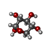  Type: D-saccharide, alpha linking / Mass: 150.130 Da / Num. of mol.: 1 / Source method: obtained synthetically / Formula: C5H10O5 / Feature type: SUBJECT OF INVESTIGATION Type: D-saccharide, alpha linking / Mass: 150.130 Da / Num. of mol.: 1 / Source method: obtained synthetically / Formula: C5H10O5 / Feature type: SUBJECT OF INVESTIGATION |
| #4: Chemical | ChemComp-YLL / (  Mass: 194.182 Da / Num. of mol.: 1 / Source method: obtained synthetically / Formula: C7H14O6 / Feature type: SUBJECT OF INVESTIGATION Mass: 194.182 Da / Num. of mol.: 1 / Source method: obtained synthetically / Formula: C7H14O6 / Feature type: SUBJECT OF INVESTIGATION |
| #5: Water | ChemComp-HOH /  Mass: 18.015 Da / Num. of mol.: 197 / Source method: isolated from a natural source / Formula: H2O Mass: 18.015 Da / Num. of mol.: 197 / Source method: isolated from a natural source / Formula: H2O |
| Has ligand of interest | Y |
| Has protein modification | Y |
-Experimental details
-Experiment
| Experiment | Method: X-RAY DIFFRACTION / Number of used crystals: 1 |
|---|
- Sample preparation
| Crystal | Density Matthews: 2.13 Å3/Da / Density % sol: 42.19 % / Description: Plate clusters |
|---|---|
| Crystal grow | Temperature: 277 K / Method: vapor diffusion, sitting drop / pH: 7.5 Details: PS: 10 mg/mL in 5 mM Na-MOPS pH 7.5, 25 mM NaCl 1:1 with WS containing 0.2 M LiCl, 20% PEG3350 PH range: 5.5-7.5 |
-Data collection
| Diffraction | Mean temperature: 100 K / Serial crystal experiment: N |
|---|---|
| Diffraction source | Source: SYNCHROTRON / Site: Diamond / Beamline: I03 / Wavelength: 0.9763 Å |
| Detector | Type: DECTRIS EIGER2 XE 16M / Detector: PIXEL / Date: Apr 25, 2021 |
| Radiation | Protocol: SINGLE WAVELENGTH / Monochromatic (M) / Laue (L): M / Scattering type: x-ray |
| Radiation wavelength | Wavelength: 0.9763 Å / Relative weight: 1 |
| Reflection | Resolution: 1.67→32.92 Å / Num. obs: 32027 / % possible obs: 97.5 % / Observed criterion σ(I): 1 / Redundancy: 6.1 % / CC1/2: 0.994 / Rmerge(I) obs: 0.181 / Rpim(I) all: 0.08 / Rrim(I) all: 0.198 / Net I/σ(I): 6.6 |
| Reflection shell | Resolution: 1.67→1.7 Å / Redundancy: 6.3 % / Rmerge(I) obs: 2.556 / Mean I/σ(I) obs: 0.8 / Num. unique obs: 1580 / CC1/2: 0.341 / Rpim(I) all: 1.095 / Rrim(I) all: 2.784 / % possible all: 96.7 |
- Processing
| Software |
| ||||||||||||||||||||||||||||||||||||||||||||||||||||||||||||||||||||||||||||||||||||||||||||||||||||||||||||||||||||||||||||||||||||||||||||||||||||||
|---|---|---|---|---|---|---|---|---|---|---|---|---|---|---|---|---|---|---|---|---|---|---|---|---|---|---|---|---|---|---|---|---|---|---|---|---|---|---|---|---|---|---|---|---|---|---|---|---|---|---|---|---|---|---|---|---|---|---|---|---|---|---|---|---|---|---|---|---|---|---|---|---|---|---|---|---|---|---|---|---|---|---|---|---|---|---|---|---|---|---|---|---|---|---|---|---|---|---|---|---|---|---|---|---|---|---|---|---|---|---|---|---|---|---|---|---|---|---|---|---|---|---|---|---|---|---|---|---|---|---|---|---|---|---|---|---|---|---|---|---|---|---|---|---|---|---|---|---|---|---|---|
| Refinement | Method to determine structure: MOLECULAR REPLACEMENT Starting model: 1tvn Resolution: 1.67→32.92 Å / Cor.coef. Fo:Fc: 0.965 / Cor.coef. Fo:Fc free: 0.951 / SU B: 3.219 / SU ML: 0.099 / Cross valid method: FREE R-VALUE / ESU R: 0.112 / ESU R Free: 0.112 Details: Hydrogens have been added in their riding positions
| ||||||||||||||||||||||||||||||||||||||||||||||||||||||||||||||||||||||||||||||||||||||||||||||||||||||||||||||||||||||||||||||||||||||||||||||||||||||
| Solvent computation | Ion probe radii: 0.8 Å / Shrinkage radii: 0.8 Å / VDW probe radii: 1.2 Å / Solvent model: MASK BULK SOLVENT | ||||||||||||||||||||||||||||||||||||||||||||||||||||||||||||||||||||||||||||||||||||||||||||||||||||||||||||||||||||||||||||||||||||||||||||||||||||||
| Displacement parameters | Biso mean: 23.653 Å2
| ||||||||||||||||||||||||||||||||||||||||||||||||||||||||||||||||||||||||||||||||||||||||||||||||||||||||||||||||||||||||||||||||||||||||||||||||||||||
| Refinement step | Cycle: LAST / Resolution: 1.67→32.92 Å
| ||||||||||||||||||||||||||||||||||||||||||||||||||||||||||||||||||||||||||||||||||||||||||||||||||||||||||||||||||||||||||||||||||||||||||||||||||||||
| Refine LS restraints |
| ||||||||||||||||||||||||||||||||||||||||||||||||||||||||||||||||||||||||||||||||||||||||||||||||||||||||||||||||||||||||||||||||||||||||||||||||||||||
| LS refinement shell |
|
