- EMDB-8590: Subtomogram average of ribosome from Trypanosoma brucei -
+
Open data
ID or keywords:
Loading...
-
Basic information
Entry
Database: EMDB / ID: EMD-8590
Title
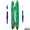
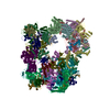
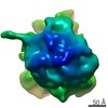
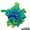
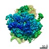
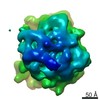
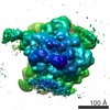
Subtomogram average of ribosome from Trypanosoma brucei
Map data
Subtomogram average of ribosome from Trypanosoma brucei
Sample
Complex: Ribosomes in cell
Function / homology
Function and homology information
organellar small ribosomal subunit / organellar large ribosomal subunit / nuclear lumen / mitochondrial large ribosomal subunit / ciliary transition zone / phosphate ion binding / negative regulation of translational frameshifting / endonucleolytic cleavage to generate mature 3'-end of SSU-rRNA from (SSU-rRNA, 5.8S rRNA, LSU-rRNA) / protein-RNA complex assembly / maturation of LSU-rRNA ...organellar small ribosomal subunit / organellar large ribosomal subunit / nuclear lumen / mitochondrial large ribosomal subunit / ciliary transition zone / phosphate ion binding / negative regulation of translational frameshifting / endonucleolytic cleavage to generate mature 3'-end of SSU-rRNA from (SSU-rRNA, 5.8S rRNA, LSU-rRNA) / protein-RNA complex assembly / maturation of LSU-rRNA / endonucleolytic cleavage in ITS1 to separate SSU-rRNA from 5.8S rRNA and LSU-rRNA from tricistronic rRNA transcript (SSU-rRNA, 5.8S rRNA, LSU-rRNA) / translation regulator activity / rescue of stalled cytosolic ribosome / protein kinase C binding / ribosome assembly / ribosomal large subunit biogenesis / maturation of LSU-rRNA from tricistronic rRNA transcript (SSU-rRNA, 5.8S rRNA, LSU-rRNA) / maturation of SSU-rRNA from tricistronic rRNA transcript (SSU-rRNA, 5.8S rRNA, LSU-rRNA) / maturation of SSU-rRNA / regulation of cell growth / small-subunit processome / maintenance of translational fidelity / modification-dependent protein catabolic process / protein tag activity / rRNA processing / regulation of cell population proliferation / ribosome biogenesis / ribosome binding / ribosomal small subunit assembly / ribosomal small subunit biogenesis / 5S rRNA binding / ribosomal large subunit assembly / small ribosomal subunit / small ribosomal subunit rRNA binding / large ribosomal subunit rRNA binding / cytosolic small ribosomal subunit / cytosolic large ribosomal subunit / cytoplasmic translation / negative regulation of translation / rRNA binding / structural constituent of ribosome / protein ubiquitination / ribosome / translation / ribonucleoprotein complex / mRNA binding / ubiquitin protein ligase binding / nucleolus / RNA binding / zinc ion binding / nucleoplasm / nucleus / cytoplasm / cytosol Similarity search - Function
Ribosomal protein L14, KOW motif / : / Ribosomal protein L28e / : / : / : / Ribosomal protein L23 / Ribosomal protein L2, archaeal-type / Ribosomal L28e/Mak16 / Ribosomal L28e protein family ...Ribosomal protein L14, KOW motif / : / Ribosomal protein L28e / : / : / : / Ribosomal protein L23 / Ribosomal protein L2, archaeal-type / Ribosomal L28e/Mak16 / Ribosomal L28e protein family / Ribosomal protein L1, conserved site / Ribosomal protein L1 signature. / Ribosomal protein L1 / : / Ribosomal protein S26e signature. / Ribosomal protein L1, 3-layer alpha/beta-sandwich / Ribosomal protein L13e, conserved site / Ribosomal protein L13e signature. / : / Ribosomal protein S12e signature. / Ribosomal protein S26e / Ribosomal protein S26e superfamily / Ribosomal protein S26e / Ribosomal protein S12e / Ribosomal protein L29e / Ribosomal L29e protein family / Small (40S) ribosomal subunit Asc1/RACK1 / Ribosomal protein L22e / Ribosomal protein L22e superfamily / Ribosomal L22e protein family / Ribosomal protein L1-like / Ribosomal protein L1/ribosomal biogenesis protein / Ribosomal protein L1p/L10e family / Ribosomal protein S21e / Ribosomal protein S5, eukaryotic/archaeal / Ribosomal protein S21e superfamily / Ribosomal protein S21e / Ribosomal protein S2, eukaryotic / Ribosomal protein L38e / Ribosomal protein L38e superfamily / Ribosomal L38e protein family / Ribosomal protein L13e / Ribosomal protein L13e / Ribosomal protein L19, eukaryotic / S27a-like superfamily / Ribosomal protein L10e, conserved site / Ribosomal protein L44e signature. / Ribosomal protein L10e signature. / 40S Ribosomal protein S10 / 60S ribosomal protein L18a/ L20, eukaryotes / Ribosomal protein L10e / Plectin/S10, N-terminal / Plectin/S10 domain / Ribosomal protein L18/L18-A/B/e, conserved site / Ribosomal protein L18e signature. / Ribosomal protein L5 eukaryotic, C-terminal / Ribosomal L18 C-terminal region / Ribosomal protein S30 / Ribosomal protein S30 / : / Ribosomal L40e family / Ribosomal protein S27a / Ribosomal protein S27a / Ribosomal protein S27a / Ribosomal protein S25 / S25 ribosomal protein / Ribosomal protein L44e / Ribosomal protein L44 / Ribosomal protein L30e signature 1. / 50S ribosomal protein L18Ae/60S ribosomal protein L20 and L18a / Ribosomal_L40e / Ribosomal protein L40e / Ribosomal protein L40e superfamily / Ribosomal protein 50S-L18Ae/60S-L20/60S-L18A / Ribosomal proteins 50S-L18Ae/60S-L20/60S-L18A / Eukaryotic Ribosomal Protein L27, KOW domain / Ribosomal protein 60S L18 and 50S L18e / Ribosomal protein L27e / Ribosomal protein L27e superfamily / Ribosomal L27e protein family / Ribosomal protein S2, eukaryotic/archaeal / : / Ribosomal Protein L6, KOW domain / 60S ribosomal protein L35 / Ribosomal protein L39e, conserved site / Ribosomal protein L39e signature. / Ribosomal protein L30e signature 2. / Ribosomal protein L30e, conserved site / : / Ribosomal protein S3, eukaryotic/archaeal / Ribosomal protein L34Ae / Ribosomal protein L34e / Ribosomal protein L7A/L8 / 60S ribosomal protein L19 / Ribosomal protein L6e / Ribosomal protein L13, eukaryotic/archaeal / 40S ribosomal protein S4, C-terminal domain / 40S ribosomal protein S4 C-terminus / 60S ribosomal protein L6E / Ribosomal protein L7, eukaryotic Similarity search - Domain/homology
60S ribosomal protein L44 / Ubiquitin-ribosomal protein eL40 fusion protein / Large ribosomal subunit protein eL18 / Large ribosomal subunit protein eL14 / 60S ribosomal protein L27, putative / 60S ribosomal protein L18a / 40S ribosomal protein S17, putative / Guanine nucleotide-binding protein beta subunit-like protein / Small ribosomal subunit protein uS2 / Large ribosomal subunit protein eL22 ...60S ribosomal protein L44 / Ubiquitin-ribosomal protein eL40 fusion protein / Large ribosomal subunit protein eL18 / Large ribosomal subunit protein eL14 / 60S ribosomal protein L27, putative / 60S ribosomal protein L18a / 40S ribosomal protein S17, putative / Guanine nucleotide-binding protein beta subunit-like protein / Small ribosomal subunit protein uS2 / Large ribosomal subunit protein eL22 / 60S ribosomal protein L29 / 60S ribosomal protein L34, putative / 40S ribosomal protein S27, putative / Ribosomal protein / 40S ribosomal protein S26 / 40S ribosomal protein S21, putative / 40S ribosomal protein S5, putative / 60S ribosomal protein L28, putative / 60S ribosomal protein L17, putative / 40S ribosomal protein S12 / 40S ribosomal protein S4 / Ribosomal protein L37 / Small ribosomal subunit protein uS5 / 40S ribosomal protein S3 / 60S ribosomal protein L6, putative / 40S ribosomal protein S24 / 60S ribosomal protein L37a, putative / 60S ribosomal protein L24, putative / 40S ribosomal protein S10, putative / 40S ribosomal protein S18, putative / 60S ribosomal protein L30 / Small ribosomal subunit protein eS1 / 60S ribosomal protein L9, putative / 40S ribosomal protein S23, putative / 40S ribosomal proteins S11, putative / 60S ribosomal protein L32, putative / 40S ribosomal protein S6 / Ribosomal protein L36, putative / Ribosomal protein L15 / 60S ribosomal protein L5, putative / 60S ribosomal protein L26, putative / 60S ribosomal subunit protein L31, putative / 60S ribosomal protein L27a / 60S ribosomal protein L23, putative / 60S ribosomal protein L10, putative / Large ribosomal subunit protein uL5 / 40S ribosomal protein S7 / Large ribosomal subunit protein eL38 / 60S ribosomal protein L35, putative / 60S ribosomal protein L21E, putative / 40S ribosomal protein S14 / 40S ribosomal protein S33, putative / 60S ribosomal protein L39, putative / 60S ribosomal protein L7, putative / 60S ribosomal protein L13 / 60S ribosomal protein L7a / 40S ribosomal protein S9, putative / 40S ribosomal protein S16, putative / 60S ribosomal protein L2, putative / Ubiquitin-ribosomal protein eL40 fusion protein / 40S ribosomal protein S15, putative / 40S ribosomal protein S25 / 60S ribosomal protein L12, putative / 40S ribosomal protein S8 / 60S ribosomal protein L23a / 60S ribosomal protein L19, putative / 40S ribosomal protein S15a, putative / Ribosomal protein L3, mitochondrial, putative / 60S ribosomal protein L4 / Ribosomal protein S19, putative / 60S ribosomal protein L35A, putative / 40S ribosomal protein S30 / 60S ribosomal protein L13a, putative / 40S ribosomal protein S13, putative Similarity search - Component
Biological species
Trypanosoma brucei (eukaryote)
Method
subtomogram averaging / cryo EM / Resolution: 38.5 Å
National Institutes of Health/National Institute of General Medical Sciences (NIH/NIGMS)
R01GM080139, P01NS092525, P41GM103832
United States
Citation
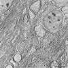
Journal: Nat Methods / Year: 2017 Title: Convolutional neural networks for automated annotation of cellular cryo-electron tomograms. Authors: Muyuan Chen / Wei Dai / Stella Y Sun / Darius Jonasch / Cynthia Y He / Michael F Schmid / Wah Chiu / Steven J Ludtke / Abstract: Cellular electron cryotomography offers researchers the ability to observe macromolecules frozen in action in situ, but a primary challenge with this technique is identifying molecular components ...Cellular electron cryotomography offers researchers the ability to observe macromolecules frozen in action in situ, but a primary challenge with this technique is identifying molecular components within the crowded cellular environment. We introduce a method that uses neural networks to dramatically reduce the time and human effort required for subcellular annotation and feature extraction. Subsequent subtomogram classification and averaging yield in situ structures of molecular components of interest. The method is available in the EMAN2.2 software package.
History
Deposition
Feb 3, 2017
-
Header (metadata) release
Mar 1, 2017
-
Map release
Mar 1, 2017
-
Update
Jan 29, 2020
-
Current status
Jan 29, 2020
Processing site: RCSB / Status: Released
-
Structure visualization
Movie
Surface view with section colored by density value
In the structure databanks used in Yorodumi, some data are registered as the other names, "COVID-19 virus" and "2019-nCoV". Here are the details of the virus and the list of structure data.
Jan 31, 2019. EMDB accession codes are about to change! (news from PDBe EMDB page)
EMDB accession codes are about to change! (news from PDBe EMDB page)
The allocation of 4 digits for EMDB accession codes will soon come to an end. Whilst these codes will remain in use, new EMDB accession codes will include an additional digit and will expand incrementally as the available range of codes is exhausted. The current 4-digit format prefixed with “EMD-” (i.e. EMD-XXXX) will advance to a 5-digit format (i.e. EMD-XXXXX), and so on. It is currently estimated that the 4-digit codes will be depleted around Spring 2019, at which point the 5-digit format will come into force.
The EM Navigator/Yorodumi systems omit the EMD- prefix.
Related info.:Q: What is EMD? / ID/Accession-code notation in Yorodumi/EM Navigator
Yorodumi is a browser for structure data from EMDB, PDB, SASBDB, etc.
This page is also the successor to EM Navigator detail page, and also detail information page/front-end page for Omokage search.
The word "yorodu" (or yorozu) is an old Japanese word meaning "ten thousand". "mi" (miru) is to see.
Related info.:EMDB / PDB / SASBDB / Comparison of 3 databanks / Yorodumi Search / Aug 31, 2016. New EM Navigator & Yorodumi / Yorodumi Papers / Jmol/JSmol / Function and homology information / Changes in new EM Navigator and Yorodumi
 Movie
Movie Controller


 Open data
 Basic information
 Map data
 Sample
 Function and homology information
 Authors
 United States, 1 items
  Citation
 Structure visualization
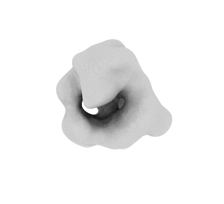
 Downloads & links
 emd_8590.png
 http://ftp.pdbj.org/pub/emdb/structures/EMD-8590


 Z (Sec.)
 Y (Row.)
 X (Col.)


 Sample components
 Processing
 Electron microscopy
 FIELD EMISSION GUN