National Institutes of Health/National Institute of General Medical Sciences (NIH/NIGMS)
R01GM080139, P01NS092525, P41GM103832
United States
Citation
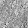
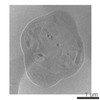
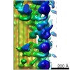
Journal: Proc Natl Acad Sci U S A / Year: 2015 Title: Electron cryotomography reveals ultrastructure alterations in platelets from patients with ovarian cancer. Authors: Rui Wang / Rebecca L Stone / Jason T Kaelber / Ryan H Rochat / Alpa M Nick / K Vinod Vijayan / Vahid Afshar-Kharghan / Michael F Schmid / Jing-Fei Dong / Anil K Sood / Wah Chiu / Abstract: Thrombocytosis and platelet hyperreactivity are known to be associated with malignancy; however, there have been no ultrastructure studies of platelets from patients with ovarian cancer. Here, we ...Thrombocytosis and platelet hyperreactivity are known to be associated with malignancy; however, there have been no ultrastructure studies of platelets from patients with ovarian cancer. Here, we used electron cryotomography (cryo-ET) to examine frozen-hydrated platelets from patients with invasive ovarian cancer (n = 12) and control subjects either with benign adnexal mass (n = 5) or free from disease (n = 6). Qualitative inspections of the tomograms indicate significant morphological differences between the cancer and control platelets, including disruption of the microtubule marginal band. Quantitative analysis of subcellular features in 120 platelet electron tomograms from these two groups showed statistically significant differences in mitochondria, as well as microtubules. These structural variations in the platelets from the patients with cancer may be correlated with the altered platelet functions associated with malignancy. Cryo-ET of platelets shows potential as a noninvasive biomarker technology for ovarian cancer and other platelet-related diseases.
History
Deposition
Feb 3, 2017
-
Header (metadata) release
Mar 1, 2017
-
Map release
Mar 1, 2017
-
Update
Jan 29, 2020
-
Current status
Jan 29, 2020
Processing site: RCSB / Status: Released
-
Structure visualization
Movie
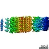
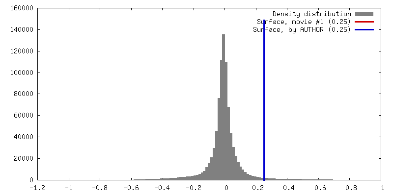
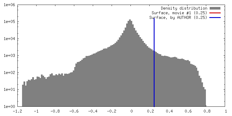
Surface view with section colored by density value
In the structure databanks used in Yorodumi, some data are registered as the other names, "COVID-19 virus" and "2019-nCoV". Here are the details of the virus and the list of structure data.
Jan 31, 2019. EMDB accession codes are about to change! (news from PDBe EMDB page)
EMDB accession codes are about to change! (news from PDBe EMDB page)
The allocation of 4 digits for EMDB accession codes will soon come to an end. Whilst these codes will remain in use, new EMDB accession codes will include an additional digit and will expand incrementally as the available range of codes is exhausted. The current 4-digit format prefixed with “EMD-” (i.e. EMD-XXXX) will advance to a 5-digit format (i.e. EMD-XXXXX), and so on. It is currently estimated that the 4-digit codes will be depleted around Spring 2019, at which point the 5-digit format will come into force.
The EM Navigator/Yorodumi systems omit the EMD- prefix.
Related info.:Q: What is EMD? / ID/Accession-code notation in Yorodumi/EM Navigator
Yorodumi is a browser for structure data from EMDB, PDB, SASBDB, etc.
This page is also the successor to EM Navigator detail page, and also detail information page/front-end page for Omokage search.
The word "yorodu" (or yorozu) is an old Japanese word meaning "ten thousand". "mi" (miru) is to see.
Related info.:EMDB / PDB / SASBDB / Comparison of 3 databanks / Yorodumi Search / Aug 31, 2016. New EM Navigator & Yorodumi / Yorodumi Papers / Jmol/JSmol / Function and homology information / Changes in new EM Navigator and Yorodumi
 Movie
Movie Controller


 Open data
 Basic information
 Map data
 Sample
 Homo sapiens (human)
 Authors
 United States, 1 items
  Citation
 Structure visualization
 Movie viewer
 Downloads & links
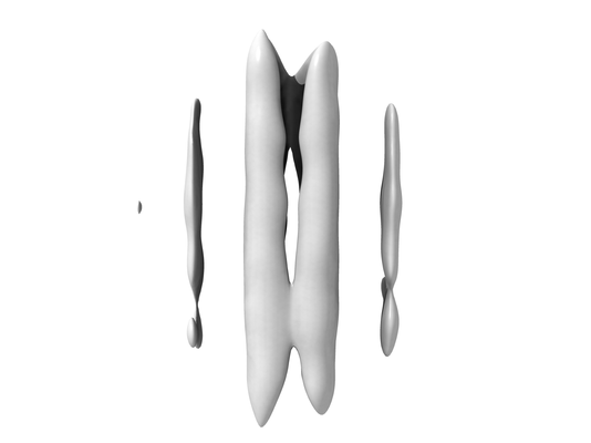 emd_8593.png
 http://ftp.pdbj.org/pub/emdb/structures/EMD-8593
 Z (Sec.)
 Y (Row.)
 X (Col.)


 Sample components
 Processing
 Electron microscopy
 FIELD EMISSION GUN