 Movie
Movie Controller

[English] 日本語
 Yorodumi
- EMDB-8528: Cryo-EM structure of 8nm repeat tubulin lattice of the ciliary mi... -
+ Open data

- Basic information
| Entry | Database: EMDB / ID: EMD-8528 | |||||||||
|---|---|---|---|---|---|---|---|---|---|---|
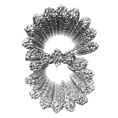
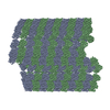
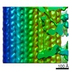
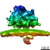
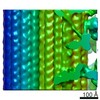
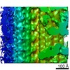
| Title | Cryo-EM structure of 8nm repeat tubulin lattice of the ciliary microtubule doublet | |||||||||
 Map data Map data | Tubulin lattice of the microtubule doublet from Tetrahymena thermophila (8nm repeat) | |||||||||
 Sample Sample |
| |||||||||
 Keywords Keywords | cilia / doublet / axoneme / tubulin / STRUCTURAL PROTEIN | |||||||||
| Function / homology |  Function and homology information Function and homology informationmicrotubule-based process / structural constituent of cytoskeleton / microtubule / Hydrolases; Acting on acid anhydrides; Acting on GTP to facilitate cellular and subcellular movement / hydrolase activity / GTPase activity / GTP binding / metal ion binding Similarity search - Function | |||||||||
| Biological species |   Tetrahymena thermophila (eukaryote) Tetrahymena thermophila (eukaryote) | |||||||||
| Method | single particle reconstruction / cryo EM / Resolution: 5.7 Å | |||||||||
 Authors Authors | Ichikawa M / Liu D | |||||||||
| Funding support |  Canada, 1 items Canada, 1 items
| |||||||||
 Citation Citation | Journal: Nat Commun / Year: 2017 Title: Subnanometre-resolution structure of the doublet microtubule reveals new classes of microtubule-associated proteins. Authors: Muneyoshi Ichikawa / Dinan Liu / Panagiotis L Kastritis / Kaustuv Basu / Tzu Chin Hsu / Shunkai Yang / Khanh Huy Bui /  Abstract: Cilia are ubiquitous, hair-like appendages found in eukaryotic cells that carry out functions of cell motility and sensory reception. Cilia contain an intriguing cytoskeletal structure, termed the ...Cilia are ubiquitous, hair-like appendages found in eukaryotic cells that carry out functions of cell motility and sensory reception. Cilia contain an intriguing cytoskeletal structure, termed the axoneme that consists of nine doublet microtubules radially interlinked and longitudinally organized in multiple specific repeat units. Little is known, however, about how the axoneme allows cilia to be both actively bendable and sturdy or how it is assembled. To answer these questions, we used cryo-electron microscopy to structurally analyse several of the repeating units of the doublet at sub-nanometre resolution. This structural detail enables us to unambiguously assign α- and β-tubulins in the doublet microtubule lattice. Our study demonstrates the existence of an inner sheath composed of different kinds of microtubule inner proteins inside the doublet that likely stabilizes the structure and facilitates the specific building of the B-tubule. | |||||||||
| History |
|
- Structure visualization
| Movie |
Movie viewer |
|---|---|
| Structure viewer | EM map: SurfViewMolmilJmol/JSmol |
| Supplemental images |
- Downloads & links
-EMDB archive
| Map data | emd_8528.map.gz | 12.6 MB | EMDB map data format | |
|---|---|---|---|---|
| Header (meta data) | emd-8528-v30.xmlemd-8528.xml | 15.7 KB 15.7 KB | Display Display | EMDB header |
| Images |  emd_8528.png emd_8528.png | 62.7 KB | ||
| Filedesc metadata | emd-8528.cif.gz | 6.1 KB | ||
| Archive directory |  http://ftp.pdbj.org/pub/emdb/structures/EMD-8528ftp://ftp.pdbj.org/pub/emdb/structures/EMD-8528 http://ftp.pdbj.org/pub/emdb/structures/EMD-8528ftp://ftp.pdbj.org/pub/emdb/structures/EMD-8528 | HTTPS FTP |
-Related structure data
| Related structure data |  5ubqMC  8532C  8537C 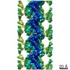 8539C 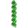 5ucyC C: citing same article ( M: atomic model generated by this map |
|---|---|
| Similar structure data |
-Links
| EMDB pages | EMDB (EBI/PDBe) / EMDataResource |
|---|---|
| Related items in Molecule of the Month |


-Map
| File | Download / File: emd_8528.map.gz / Format: CCP4 / Size: 64 MB / Type: IMAGE STORED AS FLOATING POINT NUMBER (4 BYTES) | ||||||||||||||||||||||||||||||||||||||||||||||||||||||||||||
|---|---|---|---|---|---|---|---|---|---|---|---|---|---|---|---|---|---|---|---|---|---|---|---|---|---|---|---|---|---|---|---|---|---|---|---|---|---|---|---|---|---|---|---|---|---|---|---|---|---|---|---|---|---|---|---|---|---|---|---|---|---|
| Annotation | Tubulin lattice of the microtubule doublet from Tetrahymena thermophila (8nm repeat) | ||||||||||||||||||||||||||||||||||||||||||||||||||||||||||||
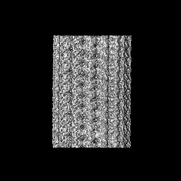
| Projections & slices | Image control
Images are generated by Spider. | ||||||||||||||||||||||||||||||||||||||||||||||||||||||||||||
| Voxel size | X=Y=Z: 2.75 Å | ||||||||||||||||||||||||||||||||||||||||||||||||||||||||||||
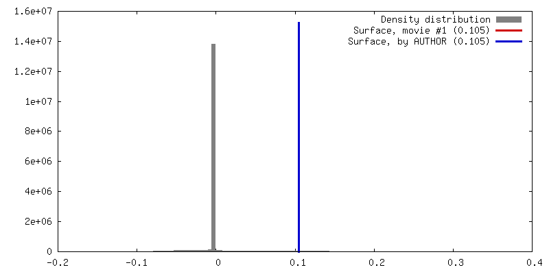
| Density |
| ||||||||||||||||||||||||||||||||||||||||||||||||||||||||||||
| Symmetry | Space group: 1 | ||||||||||||||||||||||||||||||||||||||||||||||||||||||||||||
| Details | EMDB XML:
CCP4 map header:
| ||||||||||||||||||||||||||||||||||||||||||||||||||||||||||||
 Z (Sec.)
 Y (Row.)
 X (Col.)


-Supplemental data
- Sample components
-Entire : 8nm-repeat tubulin lattice of microtubule doublet
| Entire | Name: 8nm-repeat tubulin lattice of microtubule doublet |
|---|---|
| Components |
|
-Supramolecule #1: 8nm-repeat tubulin lattice of microtubule doublet
| Supramolecule | Name: 8nm-repeat tubulin lattice of microtubule doublet / type: organelle_or_cellular_component / ID: 1 / Parent: 0 / Macromolecule list: #1-#2 |
|---|---|
| Source (natural) | Organism: Tetrahymena thermophila (eukaryote) / Strain: SB255 / Organelle: cilia / Location in cell: cilia |
-Macromolecule #1: Tubulin alpha chain
| Macromolecule | Name: Tubulin alpha chain / type: protein_or_peptide / ID: 1 / Number of copies: 3 / Enantiomer: LEVO |
|---|---|
| Source (natural) | Organism: Tetrahymena thermophila (eukaryote) |
| Molecular weight | Theoretical: 48.788254 KDa |
| Sequence | String: MREVISIHVG QGGIQVGNAC WELFCLEHGI QPDGQMPSDK TIGGGDDAFN TFFSETGAGK HVPRAVFLDL EPTVIDEVRT GTYRQLFHP EQLISGKEDA ANNFARGHYT IGKEIVDLCL DRIRKLADNC TGLQGFLVFN SVGGGTGSGL GSLLLERLSV D YGKKSKLG ...String: MREVISIHVG QGGIQVGNAC WELFCLEHGI QPDGQMPSDK TIGGGDDAFN TFFSETGAGK HVPRAVFLDL EPTVIDEVRT GTYRQLFHP EQLISGKEDA ANNFARGHYT IGKEIVDLCL DRIRKLADNC TGLQGFLVFN SVGGGTGSGL GSLLLERLSV D YGKKSKLG FTIYPSPQVS TAVVEPYNSI LSTHSLLEHT DVAVMLDNEA IYDICRRNLD IERPTYTNLN RLIAQVISSL TA SLRFDGA LNVDITEFQT NLVPYPRIHF MLSSYAPIIS AEKAYHEQLS VAEITNSAFE PANMMAKCDP RHGKYMACSM MYR GDVVPK DVNASIATIK TKRTIQFVDW CPTGFKVGIN YQPPTVVPGG DLAKVMRAVC MISNSTAIAE VFSRLDHKFD LMYA KRAFV HWYVGEGMEE GEFSEAREDL AALEKDYEEV GIETAE UniProtKB: Tubulin alpha chain |
-Macromolecule #2: Tubulin beta chain
| Macromolecule | Name: Tubulin beta chain / type: protein_or_peptide / ID: 2 / Number of copies: 3 / Enantiomer: LEVO |
|---|---|
| Source (natural) | Organism: Tetrahymena thermophila (eukaryote) |
| Molecular weight | Theoretical: 48.009199 KDa |
| Sequence | String: MREIVHIQGG QCGNQIGAKF WEVISDEHGI DPTGTYHGDS DLQLERINVY YNEATGGRYV PRAILMDLEP GTMDSVRAGP FGQLFRPDN FVFGQTGAGN NWAKGHYTEG AELIDSVLDV VRKEAEGCDC LQGFQITHSL GGGTGSGMGT LLISKVREEY P DRIMETFS ...String: MREIVHIQGG QCGNQIGAKF WEVISDEHGI DPTGTYHGDS DLQLERINVY YNEATGGRYV PRAILMDLEP GTMDSVRAGP FGQLFRPDN FVFGQTGAGN NWAKGHYTEG AELIDSVLDV VRKEAEGCDC LQGFQITHSL GGGTGSGMGT LLISKVREEY P DRIMETFS VVPSPKVSDT VVEPYNATLS VHQLVENADE CMVIDNEALY DICFRTLKLT TPTYGDLNHL VSAAMSGVTC CL RFPGQLN SDLRKLAVNL IPFPRLHFFM IGFAPLTSRG SQQYRALTVP ELTQQMFDAK NMMCAADPRH GRYLTASALF RGR MSTKEV DEQMLNVQNK NSSYFVEWIP NNIKSSICDI PPKGLKMAVT FVGNSTAIQE MFKRVAEQFT AMFRRKAFLH WYTG EGMDE MEFTEAESNM NDLVSEYQQY QDAT UniProtKB: Tubulin beta chain |
-Macromolecule #3: GUANOSINE-5'-TRIPHOSPHATE
| Macromolecule | Name: GUANOSINE-5'-TRIPHOSPHATE / type: ligand / ID: 3 / Number of copies: 3 / Formula: GTP |
|---|---|
| Molecular weight | Theoretical: 523.18 Da |
| Chemical component information |  ChemComp-GTP: |
-Macromolecule #4: MAGNESIUM ION
| Macromolecule | Name: MAGNESIUM ION / type: ligand / ID: 4 / Number of copies: 3 / Formula: MG |
|---|---|
| Molecular weight | Theoretical: 24.305 Da |
-Macromolecule #5: GUANOSINE-5'-DIPHOSPHATE
| Macromolecule | Name: GUANOSINE-5'-DIPHOSPHATE / type: ligand / ID: 5 / Number of copies: 3 / Formula: GDP |
|---|---|
| Molecular weight | Theoretical: 443.201 Da |
| Chemical component information |  ChemComp-GDP: |
-Experimental details
-Structure determination
| Method | cryo EM |
|---|---|
 Processing Processing | single particle reconstruction |
| Aggregation state | filament |
-Sample preparation
| Concentration | 2 mg/mL |
|---|---|
| Buffer | pH: 7.4 |
| Grid | Model: Quantifoil R2/2 / Material: COPPER / Mesh: 200 / Support film - Material: CARBON / Support film - topology: HOLEY / Pretreatment - Type: GLOW DISCHARGE / Pretreatment - Time: 30 sec. |
| Vitrification | Cryogen name: ETHANE / Chamber humidity: 100 % / Chamber temperature: 298 K / Instrument: FEI VITROBOT MARK IV / Details: Blot force 3 for 5 seconds. |
- Electron microscopy
| Microscope | FEI TITAN KRIOS |
|---|---|
| Image recording | Film or detector model: FEI FALCON II (4k x 4k) / Detector mode: INTEGRATING / Digitization - Frames/image: 1-7 / Number real images: 5983 / Average exposure time: 1.4 sec. / Average electron dose: 45.0 e/Å2 |
| Electron beam | Acceleration voltage: 300 kV / Electron source:  FIELD EMISSION GUN FIELD EMISSION GUN |
| Electron optics | C2 aperture diameter: 50.0 µm / Illumination mode: FLOOD BEAM / Imaging mode: BRIGHT FIELD / Cs: 2.7 mm / Nominal defocus max: 3.8000000000000003 µm / Nominal defocus min: 1.2 µm / Nominal magnification: 59000 |
| Sample stage | Specimen holder model: FEI TITAN KRIOS AUTOGRID HOLDER / Cooling holder cryogen: NITROGEN |
| Experimental equipment |  Model: Titan Krios / Image courtesy: FEI Company |
+Image processing
-Atomic model buiding 1
| Refinement | Space: REAL / Protocol: OTHER |
|---|---|
| Output model | PDB-5ubq: |
