+ Open data
- Basic information
| Entry | Database: PDB / ID: 7vlx | |||||||||
|---|---|---|---|---|---|---|---|---|---|---|
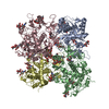
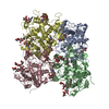
| Title | Cryo-EM structures of Listeria monocytogenes man-PTS | |||||||||
 Components Components |
| |||||||||
 Keywords Keywords | MEMBRANE PROTEIN / antimicrobial peptides / bacteriocins / pediocin PA-1 / pediocin-like/class IIa bacteriocins / antibiotic resistance / mannose phosphotransferase / man-PTS | |||||||||
| Function / homology |  Function and homology information Function and homology informationphosphoenolpyruvate-dependent sugar phosphotransferase system / membrane / plasma membrane Similarity search - Function | |||||||||
| Biological species |  Listeria monocytogenes (bacteria) Listeria monocytogenes (bacteria) | |||||||||
| Method | ELECTRON MICROSCOPY / single particle reconstruction / cryo EM / Resolution: 3.12 Å | |||||||||
 Authors Authors | Wang, J.W. | |||||||||
| Funding support |  China, 2items China, 2items
| |||||||||
 Citation Citation |  Journal: Appl Environ Microbiol / Year: 2022 Journal: Appl Environ Microbiol / Year: 2022Title: Structural Basis of Pore Formation in the Mannose Phosphotransferase System by Pediocin PA-1. Authors: Liyan Zhu / Jianwei Zeng / Chang Wang / Jiawei Wang /  Abstract: Bacteriocins are ribosomally synthesized bacterial antimicrobial peptides that have a narrow spectrum of antibacterial activity against species closely related to the producers. Pediocin-like (or ...Bacteriocins are ribosomally synthesized bacterial antimicrobial peptides that have a narrow spectrum of antibacterial activity against species closely related to the producers. Pediocin-like (or class IIa) bacteriocins (PLBs) exhibit antibacterial activity against several Gram-positive bacterial strains by forming pores in the cytoplasmic membrane of target cells with a specific receptor, the mannose phosphotransferase system (man-PTS). In this study, we report the cryo-electron microscopy structures of man-PTS from Listeria monocytogenes alone and its complex with pediocin PA-1, the first and most extensively studied representative PLB, at resolutions of 3.12 and 2.45 Å, respectively. The structures revealed that the binding of pediocin PA-1 opens the Core domain of man-PTS away from its Vmotif domain, creating a pore through the cytoplasmic membranes of target cells. During this process, the N-terminal β-sheet region of pediocin PA-1 can specifically attach to the extracellular surface of the man-PTS Core domain, whereas the C-terminal half penetrates the membrane and cracks the man-PTS like a wedge. Thus, our findings shed light on a design of novel PLBs that can kill the target pathogenic bacteria. Listeria monocytogenes is a ubiquitous microorganism responsible for listeriosis, a rare but severe disease in humans, who become infected by ingesting contaminated food products (i.e., dairy, meat, fish, and vegetables): the disease has a fatality rate of 33%. Pediocin PA-1 is an important commercial additive used in food production to inhibit species. The mannose phosphotransferase system (man-PTS) is responsible for the sensitivity of Listeria monocytogenes to pediocin PA-1. In this study, we report the cryo-EM structures of man-PTS from Listeria monocytogenes alone and its complex with pediocin PA-1 at resolutions of 3.12 and 2.45 Å, respectively. Our results facilitate the understanding of the mode of action of class IIa bacteriocins as an alternative to antibiotics. | |||||||||
| History |
|
- Structure visualization
| Movie |
 Movie viewer Movie viewer |
|---|---|
| Structure viewer | Molecule:  Molmil Molmil Jmol/JSmol Jmol/JSmol |
- Downloads & links
- Download
| PDBx/mmCIF format |  7vlx.cif.gz 7vlx.cif.gz | 272.4 KB | Display |  PDBx/mmCIF format PDBx/mmCIF format |
|---|---|---|---|---|
| PDB format |  pdb7vlx.ent.gz pdb7vlx.ent.gz | 223.6 KB | Display |  PDB format PDB format |
| PDBx/mmJSON format |  7vlx.json.gz 7vlx.json.gz | Tree view |  PDBx/mmJSON format PDBx/mmJSON format | |
| Others |  Other downloads Other downloads |
-Validation report
| Summary document |  7vlx_validation.pdf.gz 7vlx_validation.pdf.gz | 933 KB | Display |  wwPDB validaton report wwPDB validaton report |
|---|---|---|---|---|
| Full document |  7vlx_full_validation.pdf.gz 7vlx_full_validation.pdf.gz | 954.1 KB | Display | |
| Data in XML |  7vlx_validation.xml.gz 7vlx_validation.xml.gz | 52.5 KB | Display | |
| Data in CIF |  7vlx_validation.cif.gz 7vlx_validation.cif.gz | 79.9 KB | Display | |
| Arichive directory |  https://data.pdbj.org/pub/pdb/validation_reports/vl/7vlx https://data.pdbj.org/pub/pdb/validation_reports/vl/7vlx ftp://data.pdbj.org/pub/pdb/validation_reports/vl/7vlx ftp://data.pdbj.org/pub/pdb/validation_reports/vl/7vlx | HTTPS FTP |
-Related structure data
| Related structure data |  32030MC  7vlyC M: map data used to model this data C: citing same article ( |
|---|---|
| Similar structure data |
- Links
Links
- Assembly
| Deposited unit | 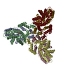
|
|---|---|
| 1 |
|
- Components
| #1: Protein | Mass: 27377.561 Da / Num. of mol.: 3 Source method: isolated from a genetically manipulated source Source: (gene. exp.)  Listeria monocytogenes (bacteria) Listeria monocytogenes (bacteria)Gene: A7N75_11070, A8L61_14515, A8N58_12480, A9809_10565, AB938_12885, ACY06_13095, AF066_07245, AF314_13905, AFT97_02015, AMC55_01930, APE37_07920, ARJ20_12560, ARS65_09750, ART25_13030, B1821_ ...Gene: A7N75_11070, A8L61_14515, A8N58_12480, A9809_10565, AB938_12885, ACY06_13095, AF066_07245, AF314_13905, AFT97_02015, AMC55_01930, APE37_07920, ARJ20_12560, ARS65_09750, ART25_13030, B1821_09270, B1N06_07640, B1N45_09425, B1N52_06240, B1N70_07565, B1O25_12365, B2H17_07955, B4960_11450, B4P04_12550, B4P23_04745, B4X79_07140, B4X80_09510, B4X87_09590, B4X92_08295, B4Y29_05710, B4Y36_09590, B4Y40_06900, B4Y49_08220, B4Y57_09590, B5K59_14490, BBW72_13535, BLH28_04840, C6R69_10580, C7H49_10125, D4920_07280, D9T24_08210, DCT16_05775, DU018_08255, E3W32_13300, E5F58_14000, E5H26_12030, EDX87_01965, EK719_12800, EPI21_11885, EXZ73_03360, F4W64_09720, F6515_08315, FA835_09550, FDP75_01900, FL871_04025, FLQ84_02200, FLQ91_10935, FLQ96_08830, FLR03_08660, FZX01_09955, G3O21_000383, GEH47_13560, GEJ92_13635, GEK33_09035, GER99_09730, GF172_05545, GGA51_12050, GHD27_05540, GIH49_05190, GIQ23_05220, GJA60_00910, GJB11_12355, GJW51_04690, GUM51_00345, GYS09_07230, GYS13_03425, GYS23_07920, GYU49_14700, GYZ23_07710, GYZ34_05285, GZI09_02550, GZI83_01895, GZP37_07990, GZS65_10905, HOY96_00900, HRK24_07015, HXD42_11005, HXF15_12125, KW30_01930, LH97_05170, LmNIHS28_00099, QU69_12075 Production host:  #2: Protein | Mass: 33402.164 Da / Num. of mol.: 3 Source method: isolated from a genetically manipulated source Source: (gene. exp.)  Listeria monocytogenes (bacteria) Listeria monocytogenes (bacteria)Gene: ARV28_12335, B4960_11455, CX098_00810, E3W32_13305, E5H26_12035, EDX87_01970, EK719_12795, EYY39_04480, F4W64_09725, FL871_04030, FZX01_09960, GEK33_09040, GH165_06065, GIH49_05195, HRK24_07020 Production host:  #3: Sugar | Has ligand of interest | Y | |
|---|
-Experimental details
-Experiment
| Experiment | Method: ELECTRON MICROSCOPY |
|---|---|
| EM experiment | Aggregation state: PARTICLE / 3D reconstruction method: single particle reconstruction |
- Sample preparation
| Component | Name: mannose-specific PTS system from Listeria monocytogenes Type: COMPLEX / Entity ID: #1-#2 / Source: RECOMBINANT |
|---|---|
| Source (natural) | Organism:  Listeria monocytogenes (bacteria) Listeria monocytogenes (bacteria) |
| Source (recombinant) | Organism:  |
| Buffer solution | pH: 7.4 |
| Specimen | Embedding applied: NO / Shadowing applied: NO / Staining applied: NO / Vitrification applied: YES |
| Vitrification | Cryogen name: NITROGEN |
- Electron microscopy imaging
| Experimental equipment |  Model: Titan Krios / Image courtesy: FEI Company |
|---|---|
| Microscopy | Model: FEI TITAN KRIOS |
| Electron gun | Electron source: LAB6 / Accelerating voltage: 300 kV / Illumination mode: FLOOD BEAM |
| Electron lens | Mode: BRIGHT FIELD |
| Image recording | Electron dose: 50 e/Å2 / Film or detector model: GATAN K3 (6k x 4k) |
- Processing
| Software | Name: PHENIX / Version: 1.19.2_4158: / Classification: refinement | ||||||||||||||||||||||||
|---|---|---|---|---|---|---|---|---|---|---|---|---|---|---|---|---|---|---|---|---|---|---|---|---|---|
| CTF correction | Type: NONE | ||||||||||||||||||||||||
| 3D reconstruction | Resolution: 3.12 Å / Resolution method: FSC 0.143 CUT-OFF / Num. of particles: 136729 / Symmetry type: POINT | ||||||||||||||||||||||||
| Refine LS restraints |
|
 Movie
Movie Controller


 UCSF Chimera




 PDBj
PDBj


