 Movie
Movie Controller

[English] 日本語
 Yorodumi
- PDB-7puy: Structure of the membrane soluble spike complex from the Lassa vi... -
+ Open data

- Basic information
| Entry | Database: PDB / ID: 7puy | ||||||
|---|---|---|---|---|---|---|---|
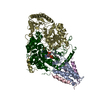
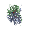
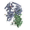
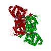
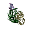
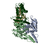
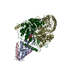
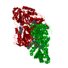
| Title | Structure of the membrane soluble spike complex from the Lassa virus in a C3-symmetric map | ||||||
 Components Components |
| ||||||
 Keywords Keywords | VIRAL PROTEIN / Spike complex / glycoprotein | ||||||
| Function / homology |  Function and homology information Function and homology informationhost cell Golgi membrane / receptor-mediated endocytosis of virus by host cell / host cell endoplasmic reticulum membrane / fusion of virus membrane with host endosome membrane / viral envelope / virion attachment to host cell / host cell plasma membrane / virion membrane / membrane / metal ion binding Similarity search - Function | ||||||
| Biological species |  Lassa virus Lassa virus | ||||||
| Method | ELECTRON MICROSCOPY / single particle reconstruction / cryo EM / Resolution: 3.3 Å | ||||||
 Authors Authors | Diskin, R. / Katz, M. | ||||||
| Funding support | 1items
| ||||||
 Citation Citation | Journal: Nature / Year: 2022 Title: Structure and receptor recognition by the Lassa virus spike complex. Authors: Michael Katz / Jonathan Weinstein / Maayan Eilon-Ashkenazy / Katrin Gehring / Hadas Cohen-Dvashi / Nadav Elad / Sarel J Fleishman / Ron Diskin /  Abstract: Lassa virus (LASV) is a human pathogen, causing substantial morbidity and mortality. Similar to other Arenaviridae, it presents a class-I spike complex on its surface that facilitates cell entry. The ...Lassa virus (LASV) is a human pathogen, causing substantial morbidity and mortality. Similar to other Arenaviridae, it presents a class-I spike complex on its surface that facilitates cell entry. The virus's cellular receptor is matriglycan, a linear carbohydrate that is present on α-dystroglycan, but the molecular mechanism that LASV uses to recognize this glycan is unknown. In addition, LASV and other arenaviruses have a unique signal peptide that forms an integral and functionally important part of the mature spike; yet the structure, function and topology of the signal peptide in the membrane remain uncertain. Here we solve the structure of a complete native LASV spike complex, finding that the signal peptide crosses the membrane once and that its amino terminus is located in the extracellular region. Together with a double-sided domain-switching mechanism, the signal peptide helps to stabilize the spike complex in its native conformation. This structure reveals that the LASV spike complex is preloaded with matriglycan, suggesting the mechanism of binding and rationalizing receptor recognition by α-dystroglycan-tropic arenaviruses. This discovery further informs us about the mechanism of viral egress and may facilitate the rational design of novel therapeutics that exploit this binding site. | ||||||
| History |
|
- Structure visualization
| Movie |
Movie viewer |
|---|---|
| Structure viewer | Molecule: MolmilJmol/JSmol |
 UCSF Chimera
- Downloads & links
-Download
| PDBx/mmCIF format | 7puy.cif.gz | 245.7 KB | Display | PDBx/mmCIF format |
|---|---|---|---|---|
| PDB format | pdb7puy.ent.gz | 193.5 KB | Display | PDB format |
| PDBx/mmJSON format | 7puy.json.gz | Tree view | PDBx/mmJSON format | |
| Others |  Other downloads Other downloads |
-Validation report
| Arichive directory | https://data.pdbj.org/pub/pdb/validation_reports/pu/7puyftp://data.pdbj.org/pub/pdb/validation_reports/pu/7puy | HTTPS FTP |
|---|
-Related structure data
| Related structure data |  13662MC  7pvdC M: map data used to model this data C: citing same article ( |
|---|---|
| Similar structure data |


-Links
 PDBj
PDBj- Assembly
| Deposited unit | 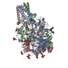
| ||||||||||||||||||||||||||||||||||||||||||||||||||||||||||||||||||||||||||||||||||||||||||||||||||||||||||||||||||||||||||||||||||||
|---|---|---|---|---|---|---|---|---|---|---|---|---|---|---|---|---|---|---|---|---|---|---|---|---|---|---|---|---|---|---|---|---|---|---|---|---|---|---|---|---|---|---|---|---|---|---|---|---|---|---|---|---|---|---|---|---|---|---|---|---|---|---|---|---|---|---|---|---|---|---|---|---|---|---|---|---|---|---|---|---|---|---|---|---|---|---|---|---|---|---|---|---|---|---|---|---|---|---|---|---|---|---|---|---|---|---|---|---|---|---|---|---|---|---|---|---|---|---|---|---|---|---|---|---|---|---|---|---|---|---|---|---|---|
| 1 |
| ||||||||||||||||||||||||||||||||||||||||||||||||||||||||||||||||||||||||||||||||||||||||||||||||||||||||||||||||||||||||||||||||||||
| Noncrystallographic symmetry (NCS) | NCS domain:
NCS domain segments:
|