 Movie
Movie Controller

[English] 日本語
 Yorodumi
- PDB-7pdh: structure of adenylyl cyclase 9 in complex with DARPin C4 and ATP-aS -
+ Open data

- Basic information
| Entry | Database: PDB / ID: 7pdh | |||||||||||||||||||||||||||||||||||||||||||||
|---|---|---|---|---|---|---|---|---|---|---|---|---|---|---|---|---|---|---|---|---|---|---|---|---|---|---|---|---|---|---|---|---|---|---|---|---|---|---|---|---|---|---|---|---|---|---|
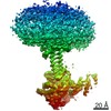
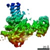
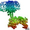
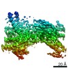
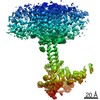
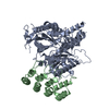
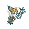
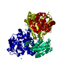
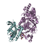
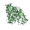
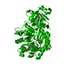
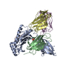
| Title | structure of adenylyl cyclase 9 in complex with DARPin C4 and ATP-aS | |||||||||||||||||||||||||||||||||||||||||||||
 Components Components |
| |||||||||||||||||||||||||||||||||||||||||||||
 Keywords Keywords | SIGNALING PROTEIN / membrane protein / adenylyl cyclase / signalling transduction. | |||||||||||||||||||||||||||||||||||||||||||||
| Function / homology |  Function and homology information Function and homology informationAdenylate cyclase activating pathway / Adenylate cyclase inhibitory pathway / PKA activation / adenylate cyclase / Hedgehog 'off' state / adenylate cyclase activity / cAMP biosynthetic process / G alpha (z) signalling events / adenylate cyclase-activating adrenergic receptor signaling pathway / adenylate cyclase-activating G protein-coupled receptor signaling pathway ...Adenylate cyclase activating pathway / Adenylate cyclase inhibitory pathway / PKA activation / adenylate cyclase / Hedgehog 'off' state / adenylate cyclase activity / cAMP biosynthetic process / G alpha (z) signalling events / adenylate cyclase-activating adrenergic receptor signaling pathway / adenylate cyclase-activating G protein-coupled receptor signaling pathway / intracellular signal transduction / ATP binding / metal ion binding / plasma membrane Similarity search - Function | |||||||||||||||||||||||||||||||||||||||||||||
| Biological species |  synthetic construct (others) | |||||||||||||||||||||||||||||||||||||||||||||
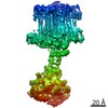
| Method | ELECTRON MICROSCOPY / single particle reconstruction / cryo EM / Resolution: 4 Å | |||||||||||||||||||||||||||||||||||||||||||||
 Authors Authors | Qi, C. / Korkhov, V.M. | |||||||||||||||||||||||||||||||||||||||||||||
| Funding support | 2items
| |||||||||||||||||||||||||||||||||||||||||||||
 Citation Citation | Journal: Nat Commun / Year: 2022 Title: Structural basis of adenylyl cyclase 9 activation. Authors: Chao Qi / Pia Lavriha / Ved Mehta / Basavraj Khanppnavar / Inayathulla Mohammed / Yong Li / Michalis Lazaratos / Jonas V Schaefer / Birgit Dreier / Andreas Plückthun / Ana-Nicoleta Bondar / ...Authors: Chao Qi / Pia Lavriha / Ved Mehta / Basavraj Khanppnavar / Inayathulla Mohammed / Yong Li / Michalis Lazaratos / Jonas V Schaefer / Birgit Dreier / Andreas Plückthun / Ana-Nicoleta Bondar / Carmen W Dessauer / Volodymyr M Korkhov /    Abstract: Adenylyl cyclase 9 (AC9) is a membrane-bound enzyme that converts ATP into cAMP. The enzyme is weakly activated by forskolin, fully activated by the G protein Gαs subunit and is autoinhibited by the ...Adenylyl cyclase 9 (AC9) is a membrane-bound enzyme that converts ATP into cAMP. The enzyme is weakly activated by forskolin, fully activated by the G protein Gαs subunit and is autoinhibited by the AC9 C-terminus. Although our recent structural studies of the AC9-Gαs complex provided the framework for understanding AC9 autoinhibition, the conformational changes that AC9 undergoes in response to activator binding remains poorly understood. Here, we present the cryo-EM structures of AC9 in several distinct states: (i) AC9 bound to a nucleotide inhibitor MANT-GTP, (ii) bound to an artificial activator (DARPin C4) and MANT-GTP, (iii) bound to DARPin C4 and a nucleotide analogue ATPαS, (iv) bound to Gαs and MANT-GTP. The artificial activator DARPin C4 partially activates AC9 by binding at a site that overlaps with the Gαs binding site. Together with the previously observed occluded and forskolin-bound conformations, structural comparisons of AC9 in the four conformations described here show that secondary structure rearrangements in the region surrounding the forskolin binding site are essential for AC9 activation. | |||||||||||||||||||||||||||||||||||||||||||||
| History |
|
- Structure visualization
| Movie |
Movie viewer |
|---|---|
| Structure viewer | Molecule: MolmilJmol/JSmol |
- Downloads & links
-Download
| PDBx/mmCIF format | 7pdh.cif.gz | 132 KB | Display | PDBx/mmCIF format |
|---|---|---|---|---|
| PDB format | pdb7pdh.ent.gz | 90.4 KB | Display | PDB format |
| PDBx/mmJSON format | 7pdh.json.gz | Tree view | PDBx/mmJSON format | |
| Others |  Other downloads Other downloads |
-Validation report
| Arichive directory | https://data.pdbj.org/pub/pdb/validation_reports/pd/7pdhftp://data.pdbj.org/pub/pdb/validation_reports/pd/7pdh | HTTPS FTP |
|---|
-Related structure data
| Related structure data | 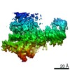 13338MC 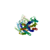 7pd4C 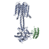 7pd8C 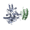 7pddC 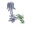 7pdeC 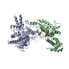 7pdfC 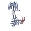 7pdgC C: citing same article ( M: map data used to model this data |
|---|---|
| Similar structure data |


-Links
 PDBj
PDBj


- Assembly
| Deposited unit | 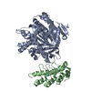
|
|---|---|
| 1 |
|
-Components
| #1: Protein | Mass: 151142.875 Da / Num. of mol.: 1 Source method: isolated from a genetically manipulated source Source: (gene. exp.)  Homo sapiens (human) / References: UniProt: E1BM79 Homo sapiens (human) / References: UniProt: E1BM79 |
|---|---|
| #2: Protein | Mass: 16042.830 Da / Num. of mol.: 1 Source method: isolated from a genetically manipulated source Source: (gene. exp.) synthetic construct (others) / Production host:  |
| Has protein modification | N |
-Experimental details
-Experiment
| Experiment | Method: ELECTRON MICROSCOPY |
|---|---|
| EM experiment | Aggregation state: PARTICLE / 3D reconstruction method: single particle reconstruction |
- Sample preparation
| Component |
| ||||||||||||||||||||||||
|---|---|---|---|---|---|---|---|---|---|---|---|---|---|---|---|---|---|---|---|---|---|---|---|---|---|
| Source (natural) |
| ||||||||||||||||||||||||
| Source (recombinant) |
| ||||||||||||||||||||||||
| Buffer solution | pH: 8 | ||||||||||||||||||||||||
| Specimen | Embedding applied: NO / Shadowing applied: NO / Staining applied: NO / Vitrification applied: YES | ||||||||||||||||||||||||
| Vitrification | Cryogen name: ETHANE |
- Electron microscopy imaging
| Experimental equipment |  Model: Titan Krios / Image courtesy: FEI Company |
|---|---|
| Microscopy | Model: FEI TITAN KRIOS |
| Electron gun | Electron source:  FIELD EMISSION GUN / Accelerating voltage: 300 kV / Illumination mode: FLOOD BEAM FIELD EMISSION GUN / Accelerating voltage: 300 kV / Illumination mode: FLOOD BEAM |
| Electron lens | Mode: BRIGHT FIELD |
| Image recording | Electron dose: 48 e/Å2 / Film or detector model: GATAN K3 (6k x 4k) |
- Processing
| Software | Name: PHENIX / Version: 1.19.2_4158: / Classification: refinement | ||||||||||||||||||||||||
|---|---|---|---|---|---|---|---|---|---|---|---|---|---|---|---|---|---|---|---|---|---|---|---|---|---|
| EM software | Name: PHENIX / Category: model refinement | ||||||||||||||||||||||||
| CTF correction | Type: NONE | ||||||||||||||||||||||||
| 3D reconstruction | Resolution: 4 Å / Resolution method: FSC 0.143 CUT-OFF / Num. of particles: 170731 / Symmetry type: POINT | ||||||||||||||||||||||||
| Refine LS restraints |
|
