 ムービー
ムービー コントローラー


+ データを開く

- 基本情報
基本情報
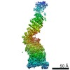
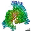
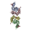
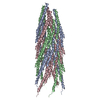
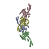
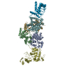
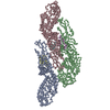
| 登録情報 | データベース: PDB / ID: 7nj1 | |||||||||
|---|---|---|---|---|---|---|---|---|---|---|
| タイトル | CryoEM structure of the human Separase-Securin complex | |||||||||
 要素 要素 |
| |||||||||
 キーワード キーワード | HYDROLASE / pseudosubstrate HEAT repeat caspase cell cycle | |||||||||
| 機能・相同性 |  機能・相同性情報 機能・相同性情報negative regulation of mitotic sister chromatid separation / negative regulation of sister chromatid cohesion / separase / meiotic chromosome separation / establishment of mitotic spindle localization / homologous chromosome segregation / meiotic spindle organization / positive regulation of mitotic metaphase/anaphase transition / cysteine-type endopeptidase inhibitor activity / mitotic sister chromatid segregation ...negative regulation of mitotic sister chromatid separation / negative regulation of sister chromatid cohesion / separase / meiotic chromosome separation / establishment of mitotic spindle localization / homologous chromosome segregation / meiotic spindle organization / positive regulation of mitotic metaphase/anaphase transition / cysteine-type endopeptidase inhibitor activity / mitotic sister chromatid segregation / mitotic cytokinesis / chromosome organization / catalytic activity / cysteine-type peptidase activity / APC/C:Cdc20 mediated degradation of Securin / molecular function activator activity / APC/C:Cdh1 mediated degradation of Cdc20 and other APC/C:Cdh1 targeted proteins in late mitosis/early G1 / mitotic spindle / SH3 domain binding / Separation of Sister Chromatids / spermatogenesis / cell division / cysteine-type endopeptidase activity / DNA repair / centrosome / apoptotic process / proteolysis / nucleus / cytoplasm / cytosol 類似検索 - 分子機能 | |||||||||
| 生物種 |  Homo sapiens (ヒト) Homo sapiens (ヒト) | |||||||||
| 手法 | 電子顕微鏡法 / 単粒子再構成法 / クライオ電子顕微鏡法 / 解像度: 2.9 Å | |||||||||
 データ登録者 データ登録者 | Yu, J. / Raia, P. / Ghent, C.M. / Raisch, T. / Sadian, Y. / Barford, D. / Raunser, S. / Morgan, D.O. / Boland, A. | |||||||||
| 資金援助 |  スイス, スイス,  米国, 2件 米国, 2件
| |||||||||
 引用 引用 | ジャーナル: Nature / 年: 2021 タイトル: Structural basis of human separase regulation by securin and CDK1-cyclin B1. 著者: Jun Yu / Pierre Raia / Chloe M Ghent / Tobias Raisch / Yashar Sadian / Simone Cavadini / Pramod M Sabale / David Barford / Stefan Raunser / David O Morgan / Andreas Boland /   要旨: In early mitosis, the duplicated chromosomes are held together by the ring-shaped cohesin complex. Separation of chromosomes during anaphase is triggered by separase-a large cysteine endopeptidase ...In early mitosis, the duplicated chromosomes are held together by the ring-shaped cohesin complex. Separation of chromosomes during anaphase is triggered by separase-a large cysteine endopeptidase that cleaves the cohesin subunit SCC1 (also known as RAD21). Separase is activated by degradation of its inhibitors, securin and cyclin B, but the molecular mechanisms of separase regulation are not clear. Here we used cryogenic electron microscopy to determine the structures of human separase in complex with either securin or CDK1-cyclin B1-CKS1. In both complexes, separase is inhibited by pseudosubstrate motifs that block substrate binding at the catalytic site and at nearby docking sites. As in Caenorhabditis elegans and yeast, human securin contains its own pseudosubstrate motifs. By contrast, CDK1-cyclin B1 inhibits separase by deploying pseudosubstrate motifs from intrinsically disordered loops in separase itself. One autoinhibitory loop is oriented by CDK1-cyclin B1 to block the catalytic sites of both separase and CDK1. Another autoinhibitory loop blocks substrate docking in a cleft adjacent to the separase catalytic site. A third separase loop contains a phosphoserine that promotes complex assembly by binding to a conserved phosphate-binding pocket in cyclin B1. Our study reveals the diverse array of mechanisms by which securin and CDK1-cyclin B1 bind and inhibit separase, providing the molecular basis for the robust control of chromosome segregation. | |||||||||
| 履歴 |
|
- 構造の表示
構造の表示
| ムービー |
ムービービューア |
|---|---|
| 構造ビューア | 分子: MolmilJmol/JSmol |
- ダウンロードとリンク
-ダウンロード
| PDBx/mmCIF形式 | 7nj1.cif.gz | 272.1 KB | 表示 | PDBx/mmCIF形式 |
|---|---|---|---|---|
| PDB形式 | pdb7nj1.ent.gz | 210.8 KB | 表示 | PDB形式 |
| PDBx/mmJSON形式 | 7nj1.json.gz | ツリー表示 | PDBx/mmJSON形式 | |
| その他 |  その他のダウンロード その他のダウンロード |
-検証レポート
| 文書・要旨 | 7nj1_validation.pdf.gz | 652.5 KB | 表示 | wwPDB検証レポート |
|---|---|---|---|---|
| 文書・詳細版 | 7nj1_full_validation.pdf.gz | 667 KB | 表示 | |
| XML形式データ | 7nj1_validation.xml.gz | 43.6 KB | 表示 | |
| CIF形式データ | 7nj1_validation.cif.gz | 65.6 KB | 表示 | |
| アーカイブディレクトリ | https://data.pdbj.org/pub/pdb/validation_reports/nj/7nj1ftp://data.pdbj.org/pub/pdb/validation_reports/nj/7nj1 | HTTPS FTP |
-関連構造データ


-リンク
 PDBj
PDBj


- 集合体
集合体
| 登録構造単位 | 
|
|---|---|
| 1 |
|
-要素
| #1: タンパク質 | 分子量: 237610.469 Da / 分子数: 1 / 由来タイプ: 組換発現 / 由来: (組換発現) Homo sapiens (ヒト) / 遺伝子: ESPL1, ESP1, KIAA0165発現宿主:   Spodoptera frugiperda (ツマジロクサヨトウ) Spodoptera frugiperda (ツマジロクサヨトウ)参照: UniProt: Q14674, separase |
|---|---|
| #2: タンパク質 | 分子量: 22052.340 Da / 分子数: 1 / 由来タイプ: 組換発現 / 由来: (組換発現) Homo sapiens (ヒト) / 遺伝子: PTTG1, EAP1, PTTG, TUTR1発現宿主: Spodoptera frugiperda (ツマジロクサヨトウ)参照: UniProt: O95997 |
-実験情報
-実験
| 実験 | 手法: 電子顕微鏡法 |
|---|---|
| EM実験 | 試料の集合状態: PARTICLE / 3次元再構成法: 単粒子再構成法 |
- 試料調製
試料調製
| 構成要素 | 名称: Inhibitory complex of human separase bound to securin. タイプ: COMPLEX / Entity ID: all / 由来: RECOMBINANT |
|---|---|
| 分子量 | 実験値: NO |
| 由来(天然) | 生物種: Homo sapiens (ヒト) |
| 由来(組換発現) | 生物種: Spodoptera frugiperda (ツマジロクサヨトウ) |
| 緩衝液 | pH: 7.8 |
| 試料 | 濃度: 0.025 mg/ml / 包埋: NO / シャドウイング: NO / 染色: NO / 凍結: YES 詳細: The sample was monodisperse. We use graphene oxide-coated EM grids. |
| 試料支持 | グリッドの材料: GOLD / グリッドのサイズ: 300 divisions/in. / グリッドのタイプ: Quantifoil R1.2/1.3 |
| 急速凍結 | 装置: LEICA EM GP / 凍結剤: ETHANE / 湿度: 90 % / 凍結前の試料温度: 293 K |
- 電子顕微鏡撮影
| 実験機器 |  モデル: Titan Krios / 画像提供: FEI Company |
|---|---|
| 顕微鏡 | モデル: FEI TITAN KRIOS |
| 電子銃 | 電子線源:  FIELD EMISSION GUN / 加速電圧: 300 kV / 照射モード: FLOOD BEAM FIELD EMISSION GUN / 加速電圧: 300 kV / 照射モード: FLOOD BEAM |
| 電子レンズ | モード: BRIGHT FIELD / 倍率(公称値): 105000 X / 最大 デフォーカス(公称値): 2500 nm / 最小 デフォーカス(公称値): 1300 nm / Calibrated defocus min: 1300 nm / 最大 デフォーカス(補正後): 2500 nm / C2レンズ絞り径: 50 µm / アライメント法: COMA FREE |
| 試料ホルダ | 凍結剤: NITROGEN 試料ホルダーモデル: FEI TITAN KRIOS AUTOGRID HOLDER |
| 撮影 | 平均露光時間: 3 sec. / 電子線照射量: 67 e/Å2 フィルム・検出器のモデル: GATAN K3 BIOQUANTUM (6k x 4k) 撮影したグリッド数: 4 / 実像数: 16540 |
- 解析
解析
| ソフトウェア | 名称: PHENIX / バージョン: 1.18rc5_3822: / 分類: 精密化 | ||||||||||||||||||||||||
|---|---|---|---|---|---|---|---|---|---|---|---|---|---|---|---|---|---|---|---|---|---|---|---|---|---|
| EMソフトウェア |
| ||||||||||||||||||||||||
| CTF補正 | タイプ: PHASE FLIPPING AND AMPLITUDE CORRECTION | ||||||||||||||||||||||||
| 対称性 | 点対称性: C1 (非対称) | ||||||||||||||||||||||||
| 3次元再構成 | 解像度: 2.9 Å / 解像度の算出法: FSC 0.143 CUT-OFF / 粒子像の数: 205300 / 対称性のタイプ: POINT | ||||||||||||||||||||||||
| 原子モデル構築 | プロトコル: AB INITIO MODEL | ||||||||||||||||||||||||
| 拘束条件 |
|
