[English] 日本語
 Yorodumi
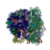
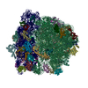
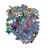
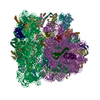
- PDB-7nhk: LsaA, an antibiotic resistance ABCF, in complex with 70S ribosome... -
+ Open data
- Basic information
| Entry | Database: PDB / ID: 7nhk | |||||||||||||||||||||
|---|---|---|---|---|---|---|---|---|---|---|---|---|---|---|---|---|---|---|---|---|---|---|
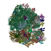
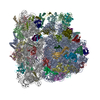
| Title | LsaA, an antibiotic resistance ABCF, in complex with 70S ribosome from Enterococcus faecalis | |||||||||||||||||||||
 Components Components |
| |||||||||||||||||||||
 Keywords Keywords | RIBOSOME / Antibiotic resistance element / ribosomal protein / LsaA / ABCF / target protection / antibiotic resistance | |||||||||||||||||||||
| Function / homology |  Function and homology information Function and homology informationregulation of translation / large ribosomal subunit / ribosome biogenesis / transferase activity / ribosomal small subunit biogenesis / ribosomal small subunit assembly / 5S rRNA binding / ribosomal large subunit assembly / small ribosomal subunit / small ribosomal subunit rRNA binding ...regulation of translation / large ribosomal subunit / ribosome biogenesis / transferase activity / ribosomal small subunit biogenesis / ribosomal small subunit assembly / 5S rRNA binding / ribosomal large subunit assembly / small ribosomal subunit / small ribosomal subunit rRNA binding / large ribosomal subunit rRNA binding / cytosolic small ribosomal subunit / cytosolic large ribosomal subunit / cytoplasmic translation / tRNA binding / negative regulation of translation / rRNA binding / structural constituent of ribosome / ribosome / translation / ribonucleoprotein complex / mRNA binding / ATP hydrolysis activity / RNA binding / zinc ion binding / ATP binding / cytosol / cytoplasm Similarity search - Function | |||||||||||||||||||||
| Biological species |  | |||||||||||||||||||||
| Method | ELECTRON MICROSCOPY / single particle reconstruction / cryo EM / Resolution: 2.9 Å | |||||||||||||||||||||
 Authors Authors | Crowe-McAuliffe, C. / Kasari, M. / Hauryliuk, V.H. / Wilson, D.N. | |||||||||||||||||||||
| Funding support |  Germany, Germany,  Sweden, Sweden,  Estonia, 6items Estonia, 6items
| |||||||||||||||||||||
 Citation Citation |  Journal: Nat Commun / Year: 2021 Journal: Nat Commun / Year: 2021Title: Structural basis of ABCF-mediated resistance to pleuromutilin, lincosamide, and streptogramin A antibiotics in Gram-positive pathogens. Authors: Caillan Crowe-McAuliffe / Victoriia Murina / Kathryn Jane Turnbull / Marje Kasari / Merianne Mohamad / Christine Polte / Hiraku Takada / Karolis Vaitkevicius / Jörgen Johansson / Zoya ...Authors: Caillan Crowe-McAuliffe / Victoriia Murina / Kathryn Jane Turnbull / Marje Kasari / Merianne Mohamad / Christine Polte / Hiraku Takada / Karolis Vaitkevicius / Jörgen Johansson / Zoya Ignatova / Gemma C Atkinson / Alex J O'Neill / Vasili Hauryliuk / Daniel N Wilson /     Abstract: Target protection proteins confer resistance to the host organism by directly binding to the antibiotic target. One class of such proteins are the antibiotic resistance (ARE) ATP-binding cassette ...Target protection proteins confer resistance to the host organism by directly binding to the antibiotic target. One class of such proteins are the antibiotic resistance (ARE) ATP-binding cassette (ABC) proteins of the F-subtype (ARE-ABCFs), which are widely distributed throughout Gram-positive bacteria and bind the ribosome to alleviate translational inhibition from antibiotics that target the large ribosomal subunit. Here, we present single-particle cryo-EM structures of ARE-ABCF-ribosome complexes from three Gram-positive pathogens: Enterococcus faecalis LsaA, Staphylococcus haemolyticus VgaA and Listeria monocytogenes VgaL. Supported by extensive mutagenesis analysis, these structures enable a general model for antibiotic resistance mediated by these ARE-ABCFs to be proposed. In this model, ABCF binding to the antibiotic-stalled ribosome mediates antibiotic release via mechanistically diverse long-range conformational relays that converge on a few conserved ribosomal RNA nucleotides located at the peptidyltransferase center. These insights are important for the future development of antibiotics that overcome such target protection resistance mechanisms. | |||||||||||||||||||||
| History |
|
- Structure visualization
| Movie |
 Movie viewer Movie viewer |
|---|---|
| Structure viewer | Molecule:  Molmil Molmil Jmol/JSmol Jmol/JSmol |
- Downloads & links
- Download
| PDBx/mmCIF format |  7nhk.cif.gz 7nhk.cif.gz | 3.2 MB | Display |  PDBx/mmCIF format PDBx/mmCIF format |
|---|---|---|---|---|
| PDB format |  pdb7nhk.ent.gz pdb7nhk.ent.gz | Display |  PDB format PDB format | |
| PDBx/mmJSON format |  7nhk.json.gz 7nhk.json.gz | Tree view |  PDBx/mmJSON format PDBx/mmJSON format | |
| Others |  Other downloads Other downloads |
-Validation report
| Summary document |  7nhk_validation.pdf.gz 7nhk_validation.pdf.gz | 1.9 MB | Display |  wwPDB validaton report wwPDB validaton report |
|---|---|---|---|---|
| Full document |  7nhk_full_validation.pdf.gz 7nhk_full_validation.pdf.gz | 2 MB | Display | |
| Data in XML |  7nhk_validation.xml.gz 7nhk_validation.xml.gz | 214.9 KB | Display | |
| Data in CIF |  7nhk_validation.cif.gz 7nhk_validation.cif.gz | 376.6 KB | Display | |
| Arichive directory |  https://data.pdbj.org/pub/pdb/validation_reports/nh/7nhk https://data.pdbj.org/pub/pdb/validation_reports/nh/7nhk ftp://data.pdbj.org/pub/pdb/validation_reports/nh/7nhk ftp://data.pdbj.org/pub/pdb/validation_reports/nh/7nhk | HTTPS FTP |
-Related structure data
| Related structure data |  12331MC  7nhlC  7nhmC  7nhnC M: map data used to model this data C: citing same article ( |
|---|---|
| Similar structure data | |
| EM raw data |  EMPIAR-10682 (Title: Affinity-purified LsaA in complex with 70S ribosomes from Enterococcus faecalis EMPIAR-10682 (Title: Affinity-purified LsaA in complex with 70S ribosomes from Enterococcus faecalisData size: 82.3 Data #1: Unaligned multi-frame micrographs of LsaA bound to 70S ribosome from Entrococcus faecalis [micrographs - multiframe]) |
- Links
Links
- Assembly
| Deposited unit | 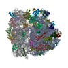
|
|---|---|
| 1 |
|
- Components
-Protein , 1 types, 1 molecules 0
| #1: Protein | Mass: 62372.363 Da / Num. of mol.: 1 / Mutation: E142Q, E452Q Source method: isolated from a genetically manipulated source Source: (gene. exp.)   |
|---|
+50S ribosomal protein ... , 28 types, 28 molecules 1245678GHIJKMNOPQRSTUVWXYZF3
-RNA chain , 5 types, 5 molecules ABaDb
| #9: RNA chain | Mass: 944000.625 Da / Num. of mol.: 1 / Source method: isolated from a natural source / Source: (natural)  |
|---|---|
| #10: RNA chain | Mass: 37433.188 Da / Num. of mol.: 1 / Source method: isolated from a natural source / Source: (natural)  |
| #30: RNA chain | Mass: 504170.562 Da / Num. of mol.: 1 / Source method: isolated from a natural source / Source: (natural)  |
| #50: RNA chain | Mass: 24811.795 Da / Num. of mol.: 1 / Source method: isolated from a natural source / Source: (natural)  |
| #51: RNA chain | Mass: 5617.279 Da / Num. of mol.: 1 / Source method: isolated from a natural source / Source: (natural)  |
-30S ribosomal protein ... , 19 types, 19 molecules cdefghijklmnopqrstu
| #31: Protein | Mass: 29500.635 Da / Num. of mol.: 1 / Source method: isolated from a natural source / Source: (natural)  |
|---|---|
| #32: Protein | Mass: 24415.184 Da / Num. of mol.: 1 / Source method: isolated from a natural source / Source: (natural)  |
| #33: Protein | Mass: 23273.652 Da / Num. of mol.: 1 / Source method: isolated from a natural source / Source: (natural)  |
| #34: Protein | Mass: 17444.357 Da / Num. of mol.: 1 / Source method: isolated from a natural source / Source: (natural)  |
| #35: Protein | Mass: 11621.188 Da / Num. of mol.: 1 / Source method: isolated from a natural source / Source: (natural)  |
| #36: Protein | Mass: 17864.625 Da / Num. of mol.: 1 / Source method: isolated from a natural source / Source: (natural)  |
| #37: Protein | Mass: 14936.396 Da / Num. of mol.: 1 / Source method: isolated from a natural source / Source: (natural)  |
| #38: Protein | Mass: 14271.480 Da / Num. of mol.: 1 / Source method: isolated from a natural source / Source: (natural)  |
| #39: Protein | Mass: 11731.739 Da / Num. of mol.: 1 / Source method: isolated from a natural source / Source: (natural)  |
| #40: Protein | Mass: 13739.913 Da / Num. of mol.: 1 / Source method: isolated from a natural source / Source: (natural)  |
| #41: Protein | Mass: 15309.817 Da / Num. of mol.: 1 / Source method: isolated from a natural source / Source: (natural)  |
| #42: Protein | Mass: 13595.774 Da / Num. of mol.: 1 / Source method: isolated from a natural source / Source: (natural)  |
| #43: Protein | Mass: 7172.593 Da / Num. of mol.: 1 / Source method: isolated from a natural source / Source: (natural)  |
| #44: Protein | Mass: 10668.236 Da / Num. of mol.: 1 / Source method: isolated from a natural source / Source: (natural)  |
| #45: Protein | Mass: 10356.150 Da / Num. of mol.: 1 / Source method: isolated from a natural source / Source: (natural)  |
| #46: Protein | Mass: 10332.100 Da / Num. of mol.: 1 / Source method: isolated from a natural source / Source: (natural)  |
| #47: Protein | Mass: 9262.891 Da / Num. of mol.: 1 / Source method: isolated from a natural source / Source: (natural)  |
| #48: Protein | Mass: 10586.332 Da / Num. of mol.: 1 / Source method: isolated from a natural source / Source: (natural)  |
| #49: Protein | Mass: 8972.320 Da / Num. of mol.: 1 / Source method: isolated from a natural source / Source: (natural)  |
-Non-polymers , 5 types, 185 molecules 


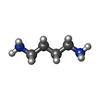


| #54: Chemical | | #55: Chemical | ChemComp-MG / #56: Chemical | #57: Chemical | ChemComp-K / #58: Chemical | ChemComp-PUT / |
|---|
-Details
| Has ligand of interest | N |
|---|
-Experimental details
-Experiment
| Experiment | Method: ELECTRON MICROSCOPY |
|---|---|
| EM experiment | Aggregation state: PARTICLE / 3D reconstruction method: single particle reconstruction |
- Sample preparation
| Component | Name: LsaA-in complex with 70S ribosome, mRNA, and tRNA / Type: RIBOSOME / Entity ID: #1-#53 / Source: NATURAL |
|---|---|
| Molecular weight | Value: 2.2 MDa / Experimental value: NO |
| Source (natural) | Organism:  |
| Buffer solution | pH: 7.5 |
| Specimen | Embedding applied: NO / Shadowing applied: NO / Staining applied: NO / Vitrification applied: YES |
| Specimen support | Grid material: COPPER / Grid mesh size: 200 divisions/in. / Grid type: Quantifoil R1.2/1.3 |
| Vitrification | Instrument: FEI VITROBOT MARK III / Cryogen name: ETHANE / Humidity: 100 % / Chamber temperature: 277.15 K / Details: 5s blotting |
- Electron microscopy imaging
| Experimental equipment |  Model: Titan Krios / Image courtesy: FEI Company |
|---|---|
| Microscopy | Model: FEI TITAN KRIOS |
| Electron gun | Electron source:  FIELD EMISSION GUN / Accelerating voltage: 300 kV / Illumination mode: FLOOD BEAM FIELD EMISSION GUN / Accelerating voltage: 300 kV / Illumination mode: FLOOD BEAM |
| Electron lens | Mode: BRIGHT FIELD / Nominal magnification: 130000 X / Nominal defocus max: -2200 nm / Nominal defocus min: -700 nm / Cs: 2.7 mm |
| Image recording | Electron dose: 38 e/Å2 / Detector mode: COUNTING / Film or detector model: GATAN K2 SUMMIT (4k x 4k) / Num. of grids imaged: 1 |
- Processing
| EM software |
| ||||||||||||
|---|---|---|---|---|---|---|---|---|---|---|---|---|---|
| CTF correction | Type: PHASE FLIPPING AND AMPLITUDE CORRECTION | ||||||||||||
| 3D reconstruction | Resolution: 2.9 Å / Resolution method: FSC 0.143 CUT-OFF / Num. of particles: 59262 / Symmetry type: POINT |
 Movie
Movie Controller




 PDBj
PDBj


