 Movie
Movie Controller

[English] 日本語
 Yorodumi
- PDB-7kog: Lethocerus Myosin II complete coiled-coil domain resolved in its ... -
+ Open data

- Basic information
| Entry | Database: PDB / ID: 7kog | ||||||||||||||||||||||||||||||||||||||||||||||||||||||
|---|---|---|---|---|---|---|---|---|---|---|---|---|---|---|---|---|---|---|---|---|---|---|---|---|---|---|---|---|---|---|---|---|---|---|---|---|---|---|---|---|---|---|---|---|---|---|---|---|---|---|---|---|---|---|---|
| Title | Lethocerus Myosin II complete coiled-coil domain resolved in its native environment | ||||||||||||||||||||||||||||||||||||||||||||||||||||||
 Components Components | Myosin heavy chain isoform Mhc_X1 | ||||||||||||||||||||||||||||||||||||||||||||||||||||||
 Keywords Keywords | MOTOR PROTEIN / striated muscle / asynchronous flight muscle | ||||||||||||||||||||||||||||||||||||||||||||||||||||||
| Biological species |  Lethocerus indicus (insect) Lethocerus indicus (insect) | ||||||||||||||||||||||||||||||||||||||||||||||||||||||
| Method | ELECTRON MICROSCOPY / single particle reconstruction / cryo EM / Resolution: 4.25 Å | ||||||||||||||||||||||||||||||||||||||||||||||||||||||
 Authors Authors | Rahmani, H. / Hu, Z. / Daneshparvar, N. / Taylor, D. / Taylor, K.A. | ||||||||||||||||||||||||||||||||||||||||||||||||||||||
| Funding support |  United States, 2items United States, 2items
| ||||||||||||||||||||||||||||||||||||||||||||||||||||||
 Citation Citation | Journal: Proc Natl Acad Sci U S A / Year: 2021 Title: The myosin II coiled-coil domain atomic structure in its native environment. Authors: Hamidreza Rahmani / Wen Ma / Zhongjun Hu / Nadia Daneshparvar / Dianne W Taylor / J Andrew McCammon / Thomas C Irving / Robert J Edwards / Kenneth A Taylor / Abstract: The atomic structure of the complete myosin tail within thick filaments isolated from flight muscle is described and compared to crystal structures of recombinant, human cardiac myosin tail segments. ...The atomic structure of the complete myosin tail within thick filaments isolated from flight muscle is described and compared to crystal structures of recombinant, human cardiac myosin tail segments. Overall, the agreement is good with three exceptions: the proximal S2, in which the filament has heads attached but the crystal structure doesn't, and skip regions 2 and 4. At the head-tail junction, the tail α-helices are asymmetrically structured encompassing well-defined unfolding of 12 residues for one myosin tail, ∼4 residues of the other, and different degrees of α-helix unwinding for both tail α-helices, thereby providing an atomic resolution description of coiled-coil "uncoiling" at the head-tail junction. Asymmetry is observed in the nonhelical C termini; one C-terminal segment is intercalated between ribbons of myosin tails, the other apparently terminating at Skip 4 of another myosin tail. Between skip residues, crystal and filament structures agree well. Skips 1 and 3 also agree well and show the expected α-helix unwinding and coiled-coil untwisting in response to skip residue insertion. Skips 2 and 4 are different. Skip 2 is accommodated in an unusual manner through an increase in α-helix radius and corresponding reduction in rise/residue. Skip 4 remains helical in one chain, with the other chain unfolded, apparently influenced by the acidic myosin C terminus. The atomic model may shed some light on thick filament mechanosensing and is a step in understanding the complex roles that thick filaments of all species undergo during muscle contraction. | ||||||||||||||||||||||||||||||||||||||||||||||||||||||
| History |
|
- Structure visualization
| Movie |
 Movie viewer Movie viewer |
|---|---|
| Structure viewer | Molecule: MolmilJmol/JSmol |
- Downloads & links
-Download
| PDBx/mmCIF format | 7kog.cif.gz | 414.8 KB | Display | PDBx/mmCIF format |
|---|---|---|---|---|
| PDB format | pdb7kog.ent.gz | 319.3 KB | Display | PDB format |
| PDBx/mmJSON format | 7kog.json.gz | Tree view | PDBx/mmJSON format | |
| Others |  Other downloads Other downloads |
-Validation report
| Arichive directory | https://data.pdbj.org/pub/pdb/validation_reports/ko/7kogftp://data.pdbj.org/pub/pdb/validation_reports/ko/7kog | HTTPS FTP |
|---|
-Related structure data
| Related structure data | 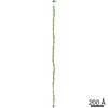 22975MC M: map data used to model this data C: citing same article ( |
|---|---|
| EM raw data | EMPIAR-10675 (Title: The Myosin II Coiled Coil Domain Atomic Structure in its Native Environment Data size: 18.8 TB / Data #1: Frame Stacks 1/3 [micrographs - multiframe] / Data #2: Frame Stacks 2/3 [micrographs - multiframe] / Data #3: Frame Stacks 3/3 [micrographs - multiframe] Data #4: Micrographs before frames alignment [micrographs - single frame] Data #5: Micrographs: frame-aligned and dose-weighted [micrographs - single frame] Data #6: Particle Stack [picked particles - single frame - unprocessed]) |
| Experimental dataset #1 | Data reference: 10.6019/EMPIAR-10675 / Data set type: EMPIAR |
-Links
 PDBj
PDBj- Assembly
| Deposited unit | 
|
|---|---|
| 1 |
|
-Components
| #1: Protein | Mass: 225227.562 Da / Num. of mol.: 2 / Source method: isolated from a natural source / Source: (natural) Lethocerus indicus (insect)Has protein modification | N | |
|---|
-Experimental details
-Experiment
| Experiment | Method: ELECTRON MICROSCOPY |
|---|---|
| EM experiment | Aggregation state: FILAMENT / 3D reconstruction method: single particle reconstruction |
- Sample preparation
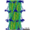
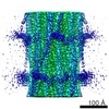
| Component | Name: Lethocerus flight muscle myosin filament / Type: ORGANELLE OR CELLULAR COMPONENT Details: The sample is a bipolar helical structure, with helical repeat 145 Angstrom and helical turn 33.98 degree. The sample has C4 symmetry. The map contains 6 unique features: myosin molecule ...Details: The sample is a bipolar helical structure, with helical repeat 145 Angstrom and helical turn 33.98 degree. The sample has C4 symmetry. The map contains 6 unique features: myosin molecule with completely resolved rods, 4 resolved non-myosin densities among the myosin rods and an annular region inside of annulus occupied by myosin rods that most likely contains paramyosin. The 4 non-myosin densities may contain parts of the proteins myofilin and flightin. Entity ID: all / Source: NATURAL |
|---|---|
| Molecular weight | Units: KILODALTONS/NANOMETER / Experimental value: NO |
| Source (natural) | Organism: Lethocerus indicus (insect) / Cellular location: myofibril / Organ: myocyte / Organelle: Sarcomere / Tissue: dorsal longitudinal indirect flight muscle |
| Buffer solution | pH: 6.8 |
| Specimen | Embedding applied: NO / Shadowing applied: NO / Staining applied: NO / Vitrification applied: YES |
| Vitrification | Cryogen name: ETHANE |
- Electron microscopy imaging
| Experimental equipment |  Model: Titan Krios / Image courtesy: FEI Company |
|---|---|
| Microscopy | Model: FEI TITAN KRIOS |
| Electron gun | Electron source:  FIELD EMISSION GUN / Accelerating voltage: 300 kV / Illumination mode: FLOOD BEAM FIELD EMISSION GUN / Accelerating voltage: 300 kV / Illumination mode: FLOOD BEAM |
| Electron lens | Mode: BRIGHT FIELD |
| Image recording | Electron dose: 60 e/Å2 / Detector mode: INTEGRATING / Film or detector model: DIRECT ELECTRON DE-64 (8k x 8k) / Num. of grids imaged: 1 / Num. of real images: 3507 |
| Image scans | Width: 8192 / Height: 8192 / Movie frames/image: 34 / Used frames/image: 1-34 |
- Processing
| Software | Name: PHENIX / Version: 1.17.1_3660: / Classification: refinement | ||||||||||||||||||||||||
|---|---|---|---|---|---|---|---|---|---|---|---|---|---|---|---|---|---|---|---|---|---|---|---|---|---|
| EM software |
| ||||||||||||||||||||||||
| CTF correction | Type: PHASE FLIPPING AND AMPLITUDE CORRECTION | ||||||||||||||||||||||||
| 3D reconstruction | Resolution: 4.25 Å / Resolution method: FSC 0.143 CUT-OFF / Num. of particles: 173515 / Symmetry type: POINT | ||||||||||||||||||||||||
| Refine LS restraints |
|
