 Movie
Movie Controller


+ Open data

- Basic information
| Entry | Database: PDB / ID: 7k00 | |||||||||
|---|---|---|---|---|---|---|---|---|---|---|
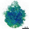
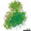
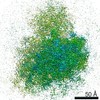
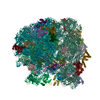
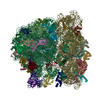
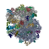
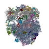
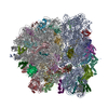
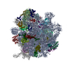
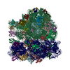
| Title | Structure of the Bacterial Ribosome at 2 Angstrom Resolution | |||||||||
 Components Components |
| |||||||||
 Keywords Keywords | RIBOSOME / antibiotics / post-translational modifications / post-transcriptional modifications | |||||||||
| Function / homology |  Function and homology information Function and homology informationpositive regulation of ribosome biogenesis / DnaA-L2 complex / negative regulation of DNA-templated DNA replication initiation / assembly of large subunit precursor of preribosome / cytosolic ribosome assembly / regulation of cell growth / mRNA 5'-UTR binding / large ribosomal subunit / small ribosomal subunit rRNA binding / ribosome binding ...positive regulation of ribosome biogenesis / DnaA-L2 complex / negative regulation of DNA-templated DNA replication initiation / assembly of large subunit precursor of preribosome / cytosolic ribosome assembly / regulation of cell growth / mRNA 5'-UTR binding / large ribosomal subunit / small ribosomal subunit rRNA binding / ribosome binding / ribosomal large subunit assembly / small ribosomal subunit / transferase activity / 5S rRNA binding / cytosolic small ribosomal subunit / cytoplasmic translation / cytosolic large ribosomal subunit / tRNA binding / rRNA binding / ribosome / structural constituent of ribosome / ribonucleoprotein complex / translation / mRNA binding / RNA binding / zinc ion binding / metal ion binding / cytosol / cytoplasm Similarity search - Function | |||||||||
| Biological species |  | |||||||||
| Method | ELECTRON MICROSCOPY / single particle reconstruction / cryo EM / Resolution: 1.98 Å | |||||||||
 Authors Authors | Watson, Z.L. / Ward, F.R. / Meheust, R. / Ad, O. / Schepartz, A. / Banfield, J.F. / Cate, J.H.D. | |||||||||
| Funding support |  United States, 2items United States, 2items
| |||||||||
 Citation Citation | Journal: Elife / Year: 2020 Title: Structure of the bacterial ribosome at 2 Å resolution. Authors: Zoe L Watson / Fred R Ward / Raphaël Méheust / Omer Ad / Alanna Schepartz / Jillian F Banfield / Jamie Hd Cate / Abstract: Using cryo-electron microscopy (cryo-EM), we determined the structure of the 70S ribosome with a global resolution of 2.0 Å. The maps reveal unambiguous positioning of protein and RNA residues, ...Using cryo-electron microscopy (cryo-EM), we determined the structure of the 70S ribosome with a global resolution of 2.0 Å. The maps reveal unambiguous positioning of protein and RNA residues, their detailed chemical interactions, and chemical modifications. Notable features include the first examples of isopeptide and thioamide backbone substitutions in ribosomal proteins, the former likely conserved in all domains of life. The maps also reveal extensive solvation of the small (30S) ribosomal subunit, and interactions with A-site and P-site tRNAs, mRNA, and the antibiotic paromomycin. The maps and models of the bacterial ribosome presented here now allow a deeper phylogenetic analysis of ribosomal components including structural conservation to the level of solvation. The high quality of the maps should enable future structural analyses of the chemical basis for translation and aid the development of robust tools for cryo-EM structure modeling and refinement. | |||||||||
| History |
|
- Structure visualization
| Movie |
Movie viewer |
|---|---|
| Structure viewer | Molecule: MolmilJmol/JSmol |
- Downloads & links
-Download
| PDBx/mmCIF format | 7k00.cif.gz | 3.3 MB | Display | PDBx/mmCIF format |
|---|---|---|---|---|
| PDB format | pdb7k00.ent.gz | Display | PDB format | |
| PDBx/mmJSON format | 7k00.json.gz | Tree view | PDBx/mmJSON format | |
| Others |  Other downloads Other downloads |
-Validation report
| Summary document | 7k00_validation.pdf.gz | 1.5 MB | Display | wwPDB validaton report |
|---|---|---|---|---|
| Full document | 7k00_full_validation.pdf.gz | 1.6 MB | Display | |
| Data in XML | 7k00_validation.xml.gz | 200.6 KB | Display | |
| Data in CIF | 7k00_validation.cif.gz | 390.9 KB | Display | |
| Arichive directory | https://data.pdbj.org/pub/pdb/validation_reports/k0/7k00ftp://data.pdbj.org/pub/pdb/validation_reports/k0/7k00 | HTTPS FTP |
-Related structure data
| Related structure data | 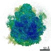 22586MC M: map data used to model this data C: citing same article ( |
|---|---|
| Similar structure data | |
| EM raw data | EMPIAR-10509 (Title: Structure of the Bacterial Ribosome at 2 Å Resolution Data size: 2.1 TB Data #1: Unaligned movies of 70S ribosome complex 1 [micrographs - multiframe] Data #2: Unaligned movies of 70S ribosome complex 2 [micrographs - multiframe]) |


-Links
 PDBj
PDBj


- Assembly
| Deposited unit | 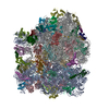
|
|---|---|
| 1 |
|
-Components
-RNA chain , 7 types, 7 molecules AabXYZ5
| #1: RNA chain | Mass: 499873.406 Da / Num. of mol.: 1 / Source method: isolated from a natural source / Source: (natural) |
|---|---|
| #22: RNA chain | Mass: 941811.562 Da / Num. of mol.: 1 / Source method: isolated from a natural source / Source: (natural) |
| #23: RNA chain | Mass: 38790.090 Da / Num. of mol.: 1 / Source method: isolated from a natural source / Source: (natural) |
| #53: RNA chain | Mass: 9099.502 Da / Num. of mol.: 1 / Source method: obtained synthetically / Source: (synth.) |
| #54: RNA chain | Mass: 24483.572 Da / Num. of mol.: 1 / Source method: obtained synthetically / Source: (synth.) |
| #55: RNA chain | Mass: 24497.602 Da / Num. of mol.: 1 / Source method: obtained synthetically / Source: (synth.) |
| #56: RNA chain | Mass: 589.430 Da / Num. of mol.: 1 / Source method: obtained synthetically / Source: (synth.) |
-30S ribosomal protein ... , 20 types, 20 molecules BCDEFGHIJKLMNOPQRSTU
| #2: Protein | Mass: 26781.670 Da / Num. of mol.: 1 / Source method: isolated from a natural source / Source: (natural) |
|---|---|
| #3: Protein | Mass: 26031.316 Da / Num. of mol.: 1 / Source method: isolated from a natural source / Source: (natural) |
| #4: Protein | Mass: 23514.199 Da / Num. of mol.: 1 / Source method: isolated from a natural source / Source: (natural) |
| #5: Protein | Mass: 17629.398 Da / Num. of mol.: 1 / Source method: isolated from a natural source / Source: (natural) |
| #6: Protein | Mass: 15727.512 Da / Num. of mol.: 1 / Source method: isolated from a natural source / Source: (natural) |
| #7: Protein | Mass: 20055.156 Da / Num. of mol.: 1 / Source method: isolated from a natural source / Source: (natural) |
| #8: Protein | Mass: 14146.557 Da / Num. of mol.: 1 / Source method: isolated from a natural source / Source: (natural) |
| #9: Protein | Mass: 14886.270 Da / Num. of mol.: 1 / Source method: isolated from a natural source / Source: (natural) |
| #10: Protein | Mass: 11755.597 Da / Num. of mol.: 1 / Source method: isolated from a natural source / Source: (natural) |
| #11: Protein | Mass: 13871.959 Da / Num. of mol.: 1 / Source method: isolated from a natural source / Source: (natural) |
| #12: Protein | Mass: 13814.249 Da / Num. of mol.: 1 / Source method: isolated from a natural source / Source: (natural) |
| #13: Protein | Mass: 13128.467 Da / Num. of mol.: 1 / Source method: isolated from a natural source / Source: (natural) |
| #14: Protein | Mass: 11606.560 Da / Num. of mol.: 1 / Source method: isolated from a natural source / Source: (natural) |
| #15: Protein | Mass: 10290.816 Da / Num. of mol.: 1 / Source method: isolated from a natural source / Source: (natural) |
| #16: Protein | Mass: 9207.572 Da / Num. of mol.: 1 / Source method: isolated from a natural source / Source: (natural) |
| #17: Protein | Mass: 9724.491 Da / Num. of mol.: 1 / Source method: isolated from a natural source / Source: (natural) |
| #18: Protein | Mass: 9005.472 Da / Num. of mol.: 1 / Source method: isolated from a natural source / Source: (natural) |
| #19: Protein | Mass: 10455.355 Da / Num. of mol.: 1 / Source method: isolated from a natural source / Source: (natural) |
| #20: Protein | Mass: 9708.464 Da / Num. of mol.: 1 / Source method: isolated from a natural source / Source: (natural) |
| #21: Protein | Mass: 8524.039 Da / Num. of mol.: 1 / Source method: isolated from a natural source / Source: (natural) |
+50S ribosomal protein ... , 29 types, 29 molecules cdefghijklmnopqrstuvwxyz01234
-Non-polymers , 6 types, 7577 molecules 


| #57: Chemical | ChemComp-PAR /  Mass: 615.628 Da / Num. of mol.: 1 / Source method: obtained synthetically / Formula: C23H45N5O14 / Comment: Antimicrobial, medication*YM Mass: 615.628 Da / Num. of mol.: 1 / Source method: obtained synthetically / Formula: C23H45N5O14 / Comment: Antimicrobial, medication*YM | ||||||||
|---|---|---|---|---|---|---|---|---|---|
| #58: Chemical | ChemComp-MG /  Mass: 24.305 Da / Num. of mol.: 309 / Source method: obtained synthetically / Formula: Mg Mass: 24.305 Da / Num. of mol.: 309 / Source method: obtained synthetically / Formula: Mg#59: Chemical | ChemComp-SPD /  Mass: 145.246 Da / Num. of mol.: 16 / Source method: obtained synthetically / Formula: C7H19N3 Mass: 145.246 Da / Num. of mol.: 16 / Source method: obtained synthetically / Formula: C7H19N3#60: Chemical | ChemComp-SPM / |  Mass: 202.340 Da / Num. of mol.: 1 / Source method: obtained synthetically / Formula: C10H26N4 Mass: 202.340 Da / Num. of mol.: 1 / Source method: obtained synthetically / Formula: C10H26N4#61: Chemical |  Mass: 65.409 Da / Num. of mol.: 2 / Source method: obtained synthetically / Formula: Zn Mass: 65.409 Da / Num. of mol.: 2 / Source method: obtained synthetically / Formula: Zn#62: Water | ChemComp-HOH / | Mass: 18.015 Da / Num. of mol.: 7248 / Source method: isolated from a natural source / Formula: H2O |
-Details
| Has ligand of interest | N |
|---|
-Experimental details
-Experiment
| Experiment | Method: ELECTRON MICROSCOPY |
|---|---|
| EM experiment | Aggregation state: PARTICLE / 3D reconstruction method: single particle reconstruction |
- Sample preparation
| Component | Name: E. coli 70S ribosome / Type: RIBOSOME Details: E. coli 70S ribosome bound to mRNA, tRNAs, and paromomycin Entity ID: #1-#56 / Source: NATURAL |
|---|---|
| Molecular weight | Value: 2.7 MDa / Experimental value: NO |
| Source (natural) | Organism: |
| Buffer solution | pH: 7.5 |
| Specimen | Conc.: 0.27 mg/ml / Embedding applied: NO / Shadowing applied: NO / Staining applied: NO / Vitrification applied: YES |
| Specimen support | Grid material: GOLD / Grid mesh size: 300 divisions/in. / Grid type: UltrAuFoil |
| Vitrification | Instrument: FEI VITROBOT MARK III / Cryogen name: ETHANE / Humidity: 100 % / Chamber temperature: 277 K |
- Electron microscopy imaging
| Experimental equipment |  Model: Titan Krios / Image courtesy: FEI Company |
|---|---|
| Microscopy | Model: FEI TITAN KRIOS |
| Electron gun | Electron source:  FIELD EMISSION GUN / Accelerating voltage: 300 kV / Illumination mode: OTHER FIELD EMISSION GUN / Accelerating voltage: 300 kV / Illumination mode: OTHER |
| Electron lens | Mode: BRIGHT FIELD / Cs: 2.7 mm |
| Image recording | Electron dose: 40 e/Å2 / Film or detector model: GATAN K3 (6k x 4k) / Num. of grids imaged: 2 |
- Processing
| EM software |
| |||||||||||||||
|---|---|---|---|---|---|---|---|---|---|---|---|---|---|---|---|---|
| CTF correction | Type: PHASE FLIPPING AND AMPLITUDE CORRECTION | |||||||||||||||
| 3D reconstruction | Resolution: 1.98 Å / Resolution method: FSC 0.143 CUT-OFF / Num. of particles: 307495 / Details: Ewald sphere corrected in RELION / Symmetry type: POINT | |||||||||||||||
| Atomic model building | PDB-ID: 4YBB Accession code: 4YBB / Source name: PDB / Type: experimental model |

