 Movie
Movie Controller

[English] 日本語
 Yorodumi
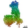
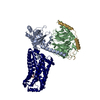
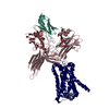
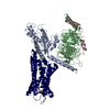
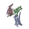
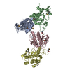
- PDB-7f6i: Cryo-EM structure of human bradykinin receptor BK2R in complex Gq... -
+ Open data

- Basic information
| Entry | Database: PDB / ID: 7f6i | |||||||||||||||||||||||||||
|---|---|---|---|---|---|---|---|---|---|---|---|---|---|---|---|---|---|---|---|---|---|---|---|---|---|---|---|---|
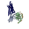
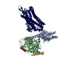
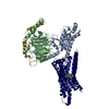
| Title | Cryo-EM structure of human bradykinin receptor BK2R in complex Gq proteins and kallidin | |||||||||||||||||||||||||||
 Components Components |
| |||||||||||||||||||||||||||
 Keywords Keywords | MEMBRANE PROTEIN / GPCR / bradykinin receptor | |||||||||||||||||||||||||||
| Function / homology |  Function and homology information Function and homology informationFatty Acids bound to GPR40 (FFAR1) regulate insulin secretion / Acetylcholine regulates insulin secretion / sensory perception of itch / phospholipase C-activating G protein-coupled glutamate receptor signaling pathway / phospholipase C-activating serotonin receptor signaling pathway / regulation of platelet activation / PLC beta mediated events / entrainment of circadian clock / regulation of canonical Wnt signaling pathway / glutamate receptor signaling pathway ...Fatty Acids bound to GPR40 (FFAR1) regulate insulin secretion / Acetylcholine regulates insulin secretion / sensory perception of itch / phospholipase C-activating G protein-coupled glutamate receptor signaling pathway / phospholipase C-activating serotonin receptor signaling pathway / regulation of platelet activation / PLC beta mediated events / entrainment of circadian clock / regulation of canonical Wnt signaling pathway / glutamate receptor signaling pathway / phototransduction, visible light / photoreceptor outer segment / postsynaptic cytosol / hormone-mediated signaling pathway / cellular response to acidic pH / Turbulent (oscillatory, disturbed) flow shear stress activates signaling by PIEZO1 and integrins in endothelial cells / mast cell degranulation / GTPase activator activity / neuropeptide signaling pathway / response to prostaglandin E / G protein-coupled receptor binding / G-protein beta/gamma-subunit complex binding / blood coagulation / Olfactory Signaling Pathway / Activation of the phototransduction cascade / G protein-coupled acetylcholine receptor signaling pathway / G beta:gamma signalling through PLC beta / Presynaptic function of Kainate receptors / Thromboxane signalling through TP receptor / Activation of G protein gated Potassium channels / Inhibition of voltage gated Ca2+ channels via Gbeta/gamma subunits / G-protein activation / Glucagon signaling in metabolic regulation / G beta:gamma signalling through CDC42 / Prostacyclin signalling through prostacyclin receptor / Synthesis, secretion, and inactivation of Glucagon-like Peptide-1 (GLP-1) / G beta:gamma signalling through BTK / photoreceptor disc membrane / ADP signalling through P2Y purinoceptor 12 / Glucagon-type ligand receptors / Sensory perception of sweet, bitter, and umami (glutamate) taste / Adrenaline,noradrenaline inhibits insulin secretion / Vasopressin regulates renal water homeostasis via Aquaporins / Glucagon-like Peptide-1 (GLP1) regulates insulin secretion / G alpha (z) signalling events / cellular response to catecholamine stimulus / ADP signalling through P2Y purinoceptor 1 / G beta:gamma signalling through PI3Kgamma / ADORA2B mediated anti-inflammatory cytokines production / adenylate cyclase-activating dopamine receptor signaling pathway / Cooperation of PDCL (PhLP1) and TRiC/CCT in G-protein beta folding / GPER1 signaling / cellular response to prostaglandin E stimulus / heterotrimeric G-protein complex / Inactivation, recovery and regulation of the phototransduction cascade / G alpha (12/13) signalling events / G-protein beta-subunit binding / extracellular vesicle / sensory perception of taste / Thrombin signalling through proteinase activated receptors (PARs) / signaling receptor complex adaptor activity / adenylate cyclase-activating G protein-coupled receptor signaling pathway / retina development in camera-type eye / fibroblast proliferation / nuclear membrane / GTPase binding / G protein activity / Ca2+ pathway / High laminar flow shear stress activates signaling by PIEZO1 and PECAM1:CDH5:KDR in endothelial cells / G alpha (i) signalling events / G alpha (s) signalling events / G alpha (q) signalling events / phospholipase C-activating G protein-coupled receptor signaling pathway / Hydrolases; Acting on acid anhydrides; Acting on GTP to facilitate cellular and subcellular movement / Ras protein signal transduction / cell population proliferation / Extra-nuclear estrogen signaling / protein stabilization / G protein-coupled receptor signaling pathway / lysosomal membrane / GTPase activity / synapse / GTP binding / protein-containing complex binding / Golgi apparatus / signal transduction / extracellular exosome / membrane / metal ion binding / plasma membrane / cytosol / cytoplasm Similarity search - Function | |||||||||||||||||||||||||||
| Biological species |  Homo sapiens (human) Homo sapiens (human) | |||||||||||||||||||||||||||
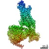
| Method | ELECTRON MICROSCOPY / single particle reconstruction / cryo EM / Resolution: 2.8 Å | |||||||||||||||||||||||||||
 Authors Authors | Shen, J. / Zhang, D. / Fu, Y. / Chen, A. / Zhang, H. | |||||||||||||||||||||||||||
| Funding support |  China, 6items China, 6items
| |||||||||||||||||||||||||||
 Citation Citation | Journal: Nat Commun / Year: 2022 Title: Cryo-EM structures of human bradykinin receptor-G proteins complexes. Authors: Jinkang Shen / Dongqi Zhang / Yao Fu / Anqi Chen / Xiaoli Yang / Haitao Zhang / Abstract: The type 2 bradykinin receptor (B2R) is a G protein-coupled receptor (GPCR) in the cardiovascular system, and the dysfunction of B2R leads to inflammation, hereditary angioedema, and pain. Bradykinin ...The type 2 bradykinin receptor (B2R) is a G protein-coupled receptor (GPCR) in the cardiovascular system, and the dysfunction of B2R leads to inflammation, hereditary angioedema, and pain. Bradykinin and kallidin are both endogenous peptide agonists of B2R, acting as vasodilators to protect the cardiovascular system. Here we determine two cryo-electron microscopy (cryo-EM) structures of human B2R-G in complex with bradykinin and kallidin at 3.0 Å and 2.9 Å resolution, respectively. The ligand-binding pocket accommodates S-shaped peptides, with aspartic acids and glutamates as an anion trap. The phenylalanines at the tail of the peptides induce significant conformational changes in the toggle switch W283, the conserved PIF, DRY, and NPxxY motifs, for the B2R activation. This further induces the extensive interactions of the intracellular loops ICL2/3 and helix 8 with G proteins. Our structures elucidate the molecular mechanisms for the ligand binding, receptor activation, and G proteins coupling of B2R. | |||||||||||||||||||||||||||
| History |
|
- Structure visualization
| Movie |
Movie viewer |
|---|---|
| Structure viewer | Molecule: MolmilJmol/JSmol |
- Downloads & links
-Download
| PDBx/mmCIF format | 7f6i.cif.gz | 191.7 KB | Display | PDBx/mmCIF format |
|---|---|---|---|---|
| PDB format | pdb7f6i.ent.gz | 140.2 KB | Display | PDB format |
| PDBx/mmJSON format | 7f6i.json.gz | Tree view | PDBx/mmJSON format | |
| Others |  Other downloads Other downloads |
-Validation report
| Arichive directory | https://data.pdbj.org/pub/pdb/validation_reports/f6/7f6iftp://data.pdbj.org/pub/pdb/validation_reports/f6/7f6i | HTTPS FTP |
|---|
-Related structure data
| Related structure data | 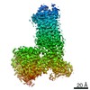 31481MC 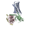 7f6hC M: map data used to model this data C: citing same article ( |
|---|---|
| Similar structure data |
-Links
 PDBj
PDBj


- Assembly
| Deposited unit | 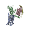
|
|---|---|
| 1 |
|
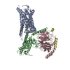
-Components
-Guanine nucleotide-binding protein ... , 3 types, 3 molecules BCD
| #3: Protein | Mass: 43534.449 Da / Num. of mol.: 1 / Mutation: R183Q, Q209L Source method: isolated from a genetically manipulated source Source: (gene. exp.) Homo sapiens (human) / Gene: GNAQ, GAQ / Production host:   Spodoptera frugiperda (fall armyworm) / References: UniProt: P50148 Spodoptera frugiperda (fall armyworm) / References: UniProt: P50148 |
|---|---|
| #4: Protein | Mass: 39086.641 Da / Num. of mol.: 1 Source method: isolated from a genetically manipulated source Source: (gene. exp.) Homo sapiens (human) / Gene: GNB1 / Production host: Spodoptera frugiperda (fall armyworm) / References: UniProt: P62873 |
| #5: Protein | Mass: 9242.612 Da / Num. of mol.: 1 Source method: isolated from a genetically manipulated source Source: (gene. exp.) Homo sapiens (human) / Gene: GNG2 / Production host: Spodoptera frugiperda (fall armyworm) / References: UniProt: P59768 |
-Protein / Protein/peptide / Non-polymers , 3 types, 5 molecules AL
| #1: Protein | Mass: 86464.734 Da / Num. of mol.: 1 Source method: isolated from a genetically manipulated source Source: (gene. exp.) Homo sapiens (human) / Production host: Spodoptera frugiperda (fall armyworm) |
|---|---|
| #2: Protein/peptide | Mass: 1191.404 Da / Num. of mol.: 1 / Source method: obtained synthetically Details: This peptide is the ligand of the protein bradykinin receptor B2R, which is the focus of research. Source: (synth.) Homo sapiens (human) |
| #6: Chemical |  Mass: 386.654 Da / Num. of mol.: 3 / Source method: obtained synthetically / Formula: C27H46O Mass: 386.654 Da / Num. of mol.: 3 / Source method: obtained synthetically / Formula: C27H46O |
-Details
| Has ligand of interest | N |
|---|---|
| Has protein modification | Y |
-Experimental details
-Experiment
| Experiment | Method: ELECTRON MICROSCOPY |
|---|---|
| EM experiment | Aggregation state: PARTICLE / 3D reconstruction method: single particle reconstruction |
- Sample preparation
| Component | Name: Bradykinin receptor BK2RR in complex with Gq proteins and kallidin Type: COMPLEX / Entity ID: #1-#5 / Source: RECOMBINANT |
|---|---|
| Molecular weight | Value: 0.173 MDa / Experimental value: YES |
| Source (natural) | Organism: Homo sapiens (human) |
| Source (recombinant) | Organism: Spodoptera frugiperda (fall armyworm) |
| Buffer solution | pH: 7.5 |
| Specimen | Embedding applied: NO / Shadowing applied: NO / Staining applied: NO / Vitrification applied: YES |
| Vitrification | Cryogen name: ETHANE |
- Electron microscopy imaging
| Experimental equipment |  Model: Titan Krios / Image courtesy: FEI Company |
|---|---|
| Microscopy | Model: FEI TITAN KRIOS |
| Electron gun | Electron source:  FIELD EMISSION GUN / Accelerating voltage: 300 kV / Illumination mode: OTHER FIELD EMISSION GUN / Accelerating voltage: 300 kV / Illumination mode: OTHER |
| Electron lens | Mode: BRIGHT FIELD |
| Image recording | Electron dose: 64 e/Å2 / Film or detector model: GATAN K2 BASE (4k x 4k) |
- Processing
| Software | Name: PHENIX / Version: 1.18.2_3874: / Classification: refinement | ||||||||||||||||||||||||
|---|---|---|---|---|---|---|---|---|---|---|---|---|---|---|---|---|---|---|---|---|---|---|---|---|---|
| EM software | Name: PHENIX / Category: model refinement | ||||||||||||||||||||||||
| CTF correction | Type: PHASE FLIPPING AND AMPLITUDE CORRECTION | ||||||||||||||||||||||||
| 3D reconstruction | Resolution: 2.8 Å / Resolution method: FSC 0.143 CUT-OFF / Num. of particles: 2042659 / Symmetry type: POINT | ||||||||||||||||||||||||
| Refine LS restraints |
|
