 Movie
Movie Controller

[English] 日本語
 Yorodumi
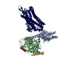
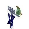
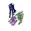
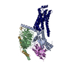
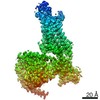
- PDB-7eib: Cryo-EM structure of the type 1 bradykinin receptor in complex wi... -
+ Open data

- Basic information
| Entry | Database: PDB / ID: 7eib | |||||||||||||||||||||
|---|---|---|---|---|---|---|---|---|---|---|---|---|---|---|---|---|---|---|---|---|---|---|
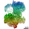
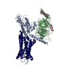
| Title | Cryo-EM structure of the type 1 bradykinin receptor in complex with the des-Arg10-kallidin and an Gq protein | |||||||||||||||||||||
 Components Components |
| |||||||||||||||||||||
 Keywords Keywords | MEMBRANE PROTEIN / Bradykinin receptors / Kinin / GPCR | |||||||||||||||||||||
| Function / homology |  Function and homology information Function and homology informationbradykinin receptor activity / positive regulation of leukocyte migration / G-protein activation / Activation of the phototransduction cascade / Glucagon-type ligand receptors / Thromboxane signalling through TP receptor / Sensory perception of sweet, bitter, and umami (glutamate) taste / G beta:gamma signalling through PI3Kgamma / G beta:gamma signalling through CDC42 / Cooperation of PDCL (PhLP1) and TRiC/CCT in G-protein beta folding ...bradykinin receptor activity / positive regulation of leukocyte migration / G-protein activation / Activation of the phototransduction cascade / Glucagon-type ligand receptors / Thromboxane signalling through TP receptor / Sensory perception of sweet, bitter, and umami (glutamate) taste / G beta:gamma signalling through PI3Kgamma / G beta:gamma signalling through CDC42 / Cooperation of PDCL (PhLP1) and TRiC/CCT in G-protein beta folding / Activation of G protein gated Potassium channels / Inhibition of voltage gated Ca2+ channels via Gbeta/gamma subunits / Ca2+ pathway / G alpha (z) signalling events / High laminar flow shear stress activates signaling by PIEZO1 and PECAM1:CDH5:KDR in endothelial cells / Glucagon-like Peptide-1 (GLP1) regulates insulin secretion / Vasopressin regulates renal water homeostasis via Aquaporins / Adrenaline,noradrenaline inhibits insulin secretion / ADP signalling through P2Y purinoceptor 12 / G alpha (q) signalling events / G alpha (i) signalling events / Thrombin signalling through proteinase activated receptors (PARs) / Activation of G protein gated Potassium channels / G-protein activation / G beta:gamma signalling through PI3Kgamma / Prostacyclin signalling through prostacyclin receptor / G beta:gamma signalling through PLC beta / ADP signalling through P2Y purinoceptor 1 / Thromboxane signalling through TP receptor / Presynaptic function of Kainate receptors / G beta:gamma signalling through CDC42 / Inhibition of voltage gated Ca2+ channels via Gbeta/gamma subunits / G alpha (12/13) signalling events / Glucagon-type ligand receptors / G beta:gamma signalling through BTK / ADP signalling through P2Y purinoceptor 12 / Adrenaline,noradrenaline inhibits insulin secretion / Cooperation of PDCL (PhLP1) and TRiC/CCT in G-protein beta folding / Ca2+ pathway / G alpha (z) signalling events / Thrombin signalling through proteinase activated receptors (PARs) / Extra-nuclear estrogen signaling / G alpha (s) signalling events / G alpha (q) signalling events / photoreceptor outer segment membrane / spectrin binding / Glucagon-like Peptide-1 (GLP1) regulates insulin secretion / G alpha (i) signalling events / High laminar flow shear stress activates signaling by PIEZO1 and PECAM1:CDH5:KDR in endothelial cells / Vasopressin regulates renal water homeostasis via Aquaporins / alkylglycerophosphoethanolamine phosphodiesterase activity / photoreceptor outer segment / response to mechanical stimulus / negative regulation of blood pressure / cardiac muscle cell apoptotic process / peptide binding / photoreceptor inner segment / positive regulation of release of sequestered calcium ion into cytosol / Peptide ligand-binding receptors / negative regulation of cell growth / cellular response to catecholamine stimulus / cell migration / adenylate cyclase-activating dopamine receptor signaling pathway / cellular response to prostaglandin E stimulus / heterotrimeric G-protein complex / G-protein beta-subunit binding / positive regulation of cytosolic calcium ion concentration / sensory perception of taste / signaling receptor complex adaptor activity / retina development in camera-type eye / cell body / GTPase binding / response to lipopolysaccharide / cellular response to hypoxia / G alpha (i) signalling events / G alpha (q) signalling events / phospholipase C-activating G protein-coupled receptor signaling pathway / cell population proliferation / inflammatory response / G protein-coupled receptor signaling pathway / GTPase activity / dendrite / synapse / protein-containing complex binding / endoplasmic reticulum / membrane / plasma membrane / cytoplasm Similarity search - Function | |||||||||||||||||||||
| Biological species |  Homo sapiens (human) Homo sapiens (human) | |||||||||||||||||||||
| Method | ELECTRON MICROSCOPY / single particle reconstruction / cryo EM / Resolution: 3 Å | |||||||||||||||||||||
 Authors Authors | Yin, Y. / Jiang, Y. | |||||||||||||||||||||
| Funding support |  China, 6items China, 6items
| |||||||||||||||||||||
 Citation Citation | Journal: Nat Struct Mol Biol / Year: 2021 Title: Molecular basis for kinin selectivity and activation of the human bradykinin receptors. Authors: Yu-Ling Yin / Chenyu Ye / Fulai Zhou / Jia Wang / Dehua Yang / Wanchao Yin / Ming-Wei Wang / H Eric Xu / Yi Jiang / Abstract: Bradykinin and kallidin are endogenous kinin peptide hormones that belong to the kallikrein-kinin system and are essential to the regulation of blood pressure, inflammation, coagulation and pain ...Bradykinin and kallidin are endogenous kinin peptide hormones that belong to the kallikrein-kinin system and are essential to the regulation of blood pressure, inflammation, coagulation and pain control. Des-Arg-kallidin, the carboxy-terminal des-Arg metabolite of kallidin, and bradykinin selectively activate two G protein-coupled receptors, type 1 and type 2 bradykinin receptors (B1R and B2R), respectively. The hyperactivation of bradykinin receptors, termed 'bradykinin storm', is associated with pulmonary edema in COVID-19 patients, suggesting that bradykinin receptors are important targets for COVID-19 intervention. Here we report two G protein-coupled complex structures of human B1R and B2R bound to des-Arg-kallidin and bradykinin, respectively. Combined with functional analysis, our structures reveal the mechanism of ligand selectivity and specific activation of the bradykinin receptor. These findings also provide a framework for guiding drug design targeting bradykinin receptors for the treatment of inflammation, cardiovascular disorders and COVID-19. | |||||||||||||||||||||
| History |
|
- Structure visualization
| Movie |
Movie viewer |
|---|---|
| Structure viewer | Molecule: MolmilJmol/JSmol |
 UCSF Chimera
- Downloads & links
-Download
| PDBx/mmCIF format | 7eib.cif.gz | 189.1 KB | Display | PDBx/mmCIF format |
|---|---|---|---|---|
| PDB format | pdb7eib.ent.gz | 136.9 KB | Display | PDB format |
| PDBx/mmJSON format | 7eib.json.gz | Tree view | PDBx/mmJSON format | |
| Others |  Other downloads Other downloads |
-Validation report
| Arichive directory | https://data.pdbj.org/pub/pdb/validation_reports/ei/7eibftp://data.pdbj.org/pub/pdb/validation_reports/ei/7eib | HTTPS FTP |
|---|
-Related structure data
| Related structure data | 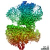 31145MC 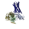 7f2oC M: map data used to model this data C: citing same article ( |
|---|---|
| Similar structure data |
-Links
 PDBj
PDBj


- Assembly
| Deposited unit | 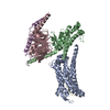
|
|---|---|
| 1 |
|
-Components
| #1: Protein | Mass: 69344.070 Da / Num. of mol.: 1 / Mutation: F126W Source method: isolated from a genetically manipulated source Source: (gene. exp.) Homo sapiens (human) / Gene: BDKRB1, BRADYB1 / Production host:   Spodoptera frugiperda (fall armyworm) / References: UniProt: P46663 Spodoptera frugiperda (fall armyworm) / References: UniProt: P46663 |
|---|---|
| #2: Protein/peptide | Mass: 1034.210 Da / Num. of mol.: 1 / Source method: obtained synthetically / Source: (synth.) Homo sapiens (human) |
| #3: Protein | Mass: 41724.383 Da / Num. of mol.: 1 Source method: isolated from a genetically manipulated source Source: (gene. exp.) Homo sapiens (human) / Production host: Spodoptera frugiperda (fall armyworm) |
| #4: Protein | Mass: 41055.867 Da / Num. of mol.: 1 Source method: isolated from a genetically manipulated source Source: (gene. exp.) Spodoptera frugiperda (fall armyworm) / References: UniProt: P54311 |
| #5: Protein | Mass: 7861.143 Da / Num. of mol.: 1 Source method: isolated from a genetically manipulated source Source: (gene. exp.) Spodoptera frugiperda (fall armyworm) / References: UniProt: P63212 |
| Has protein modification | Y |
-Experimental details
-Experiment
| Experiment | Method: ELECTRON MICROSCOPY |
|---|---|
| EM experiment | Aggregation state: PARTICLE / 3D reconstruction method: single particle reconstruction |
- Sample preparation
| Component |
| ||||||||||||||||||||||||||||||||||||||||||
|---|---|---|---|---|---|---|---|---|---|---|---|---|---|---|---|---|---|---|---|---|---|---|---|---|---|---|---|---|---|---|---|---|---|---|---|---|---|---|---|---|---|---|---|
| Source (natural) |
| ||||||||||||||||||||||||||||||||||||||||||
| Source (recombinant) |
| ||||||||||||||||||||||||||||||||||||||||||
| Buffer solution | pH: 7.5 | ||||||||||||||||||||||||||||||||||||||||||
| Specimen | Embedding applied: NO / Shadowing applied: NO / Staining applied: NO / Vitrification applied: YES | ||||||||||||||||||||||||||||||||||||||||||
| Vitrification | Cryogen name: ETHANE |
- Electron microscopy imaging
| Experimental equipment |  Model: Titan Krios / Image courtesy: FEI Company |
|---|---|
| Microscopy | Model: FEI TITAN KRIOS |
| Electron gun | Electron source: OTHER / Accelerating voltage: 300 kV / Illumination mode: OTHER |
| Electron lens | Mode: BRIGHT FIELD |
| Image recording | Electron dose: 61.8 e/Å2 / Film or detector model: OTHER |
- Processing
| Software | Name: PHENIX / Version: 1.18.2_3874: / Classification: refinement | ||||||||||||||||||||||||
|---|---|---|---|---|---|---|---|---|---|---|---|---|---|---|---|---|---|---|---|---|---|---|---|---|---|
| EM software | Name: PHENIX / Category: model refinement | ||||||||||||||||||||||||
| CTF correction | Type: NONE | ||||||||||||||||||||||||
| 3D reconstruction | Resolution: 3 Å / Resolution method: FSC 0.143 CUT-OFF / Num. of particles: 633636 / Symmetry type: POINT | ||||||||||||||||||||||||
| Refine LS restraints |
|
