 Movie
Movie Controller

[English] 日本語
 Yorodumi
- PDB-7ld4: Cryo-EM structure of the human adenosine A1 receptor-Gi2-protein ... -
+ Open data

- Basic information
| Entry | Database: PDB / ID: 7ld4 | ||||||||||||||||||||||||||||||||||||||||||||||||||||||||||||||||||||||||
|---|---|---|---|---|---|---|---|---|---|---|---|---|---|---|---|---|---|---|---|---|---|---|---|---|---|---|---|---|---|---|---|---|---|---|---|---|---|---|---|---|---|---|---|---|---|---|---|---|---|---|---|---|---|---|---|---|---|---|---|---|---|---|---|---|---|---|---|---|---|---|---|---|---|
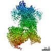
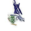
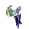
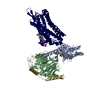
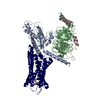
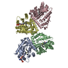
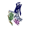
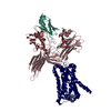
| Title | Cryo-EM structure of the human adenosine A1 receptor-Gi2-protein complex bound to its endogenous agonist | ||||||||||||||||||||||||||||||||||||||||||||||||||||||||||||||||||||||||
 Components Components |
| ||||||||||||||||||||||||||||||||||||||||||||||||||||||||||||||||||||||||
 Keywords Keywords | SIGNALING PROTEIN / membrane protein / active-state G protein-coupled receptor / adenosine A1 receptor | ||||||||||||||||||||||||||||||||||||||||||||||||||||||||||||||||||||||||
| Function / homology |  Function and homology information Function and homology informationpositive regulation of nucleoside transport / negative regulation of neurotrophin production / regulation of glomerular filtration / negative regulation of circadian sleep/wake cycle, non-REM sleep / G protein-coupled purinergic nucleotide receptor signaling pathway / negative regulation of mucus secretion / purine nucleoside binding / positive regulation of peptide secretion / negative regulation of glutamate secretion / negative regulation of synaptic transmission, GABAergic ...positive regulation of nucleoside transport / negative regulation of neurotrophin production / regulation of glomerular filtration / negative regulation of circadian sleep/wake cycle, non-REM sleep / G protein-coupled purinergic nucleotide receptor signaling pathway / negative regulation of mucus secretion / purine nucleoside binding / positive regulation of peptide secretion / negative regulation of glutamate secretion / negative regulation of synaptic transmission, GABAergic / negative regulation of long-term synaptic depression / positive regulation of dephosphorylation / negative regulation of hormone secretion / positive regulation of lipid catabolic process / mucus secretion / negative regulation of adenylate cyclase-activating adrenergic receptor signaling pathway / negative regulation of leukocyte migration / Muscarinic acetylcholine receptors / G protein-coupled acetylcholine receptor activity / regulation of sensory perception of pain / regulation of respiratory gaseous exchange by nervous system process / heterotrimeric G-protein binding / regulation of presynaptic cytosolic calcium ion concentration / negative regulation of calcium ion-dependent exocytosis / Adenosine P1 receptors / G protein-coupled adenosine receptor activity / negative regulation of adenylate cyclase activity / positive regulation of potassium ion transport / response to purine-containing compound / adenylate cyclase-inhibiting G protein-coupled acetylcholine receptor signaling pathway / G protein-coupled adenosine receptor signaling pathway / positive regulation of neural precursor cell proliferation / negative regulation of synaptic transmission / regulation of cardiac muscle cell contraction / negative regulation of systemic arterial blood pressure / long-term synaptic depression / negative regulation of synaptic transmission, glutamatergic / protein targeting to membrane / leukocyte migration / triglyceride homeostasis / presynaptic active zone / positive regulation of urine volume / gamma-aminobutyric acid signaling pathway / regulation of locomotion / negative regulation of acute inflammatory response / temperature homeostasis / positive regulation of systemic arterial blood pressure / regulation of calcium ion transport / detection of temperature stimulus involved in sensory perception of pain / negative regulation of long-term synaptic potentiation / negative regulation of apoptotic signaling pathway / fatty acid homeostasis / asymmetric synapse / axolemma / negative regulation of lipid catabolic process / G protein-coupled receptor signaling pathway, coupled to cyclic nucleotide second messenger / neuronal dense core vesicle / phagocytosis / lipid catabolic process / positive regulation of vascular associated smooth muscle cell proliferation / positive regulation of superoxide anion generation / heat shock protein binding / Adenylate cyclase inhibitory pathway / response to nutrient / calyx of Held / hippocampal mossy fiber to CA3 synapse / apoptotic signaling pathway / Regulation of insulin secretion / excitatory postsynaptic potential / G protein-coupled receptor binding / cognition / vasodilation / G-protein beta/gamma-subunit complex binding / adenylate cyclase-inhibiting G protein-coupled receptor signaling pathway / Olfactory Signaling Pathway / Activation of the phototransduction cascade / terminal bouton / G protein-coupled acetylcholine receptor signaling pathway / G beta:gamma signalling through PLC beta / Presynaptic function of Kainate receptors / Thromboxane signalling through TP receptor / Activation of G protein gated Potassium channels / Inhibition of voltage gated Ca2+ channels via Gbeta/gamma subunits / G-protein activation / Glucagon signaling in metabolic regulation / G beta:gamma signalling through CDC42 / Prostacyclin signalling through prostacyclin receptor / Synthesis, secretion, and inactivation of Glucagon-like Peptide-1 (GLP-1) / G beta:gamma signalling through BTK / photoreceptor disc membrane / ADP signalling through P2Y purinoceptor 12 / Glucagon-type ligand receptors / Sensory perception of sweet, bitter, and umami (glutamate) taste / Adrenaline,noradrenaline inhibits insulin secretion / Vasopressin regulates renal water homeostasis via Aquaporins / Glucagon-like Peptide-1 (GLP1) regulates insulin secretion / G alpha (z) signalling events / cell-cell signaling / nervous system development / cellular response to catecholamine stimulus Similarity search - Function | ||||||||||||||||||||||||||||||||||||||||||||||||||||||||||||||||||||||||
| Biological species |  Homo sapiens (human) Homo sapiens (human) | ||||||||||||||||||||||||||||||||||||||||||||||||||||||||||||||||||||||||
| Method | ELECTRON MICROSCOPY / single particle reconstruction / cryo EM / Resolution: 3.3 Å | ||||||||||||||||||||||||||||||||||||||||||||||||||||||||||||||||||||||||
 Authors Authors | Draper-Joyce, C.J. / Danev, R. / Thal, D.M. / Christopoulos, A. / Glukhova, A. | ||||||||||||||||||||||||||||||||||||||||||||||||||||||||||||||||||||||||
| Funding support |  Australia, 2items Australia, 2items
| ||||||||||||||||||||||||||||||||||||||||||||||||||||||||||||||||||||||||
 Citation Citation | Journal: Nature / Year: 2021 Title: Positive allosteric mechanisms of adenosine A receptor-mediated analgesia. Authors: Christopher J Draper-Joyce / Rebecca Bhola / Jinan Wang / Apurba Bhattarai / Anh T N Nguyen / India Cowie-Kent / Kelly O'Sullivan / Ling Yeong Chia / Hariprasad Venugopal / Celine Valant / ...Authors: Christopher J Draper-Joyce / Rebecca Bhola / Jinan Wang / Apurba Bhattarai / Anh T N Nguyen / India Cowie-Kent / Kelly O'Sullivan / Ling Yeong Chia / Hariprasad Venugopal / Celine Valant / David M Thal / Denise Wootten / Nicolas Panel / Jens Carlsson / Macdonald J Christie / Paul J White / Peter Scammells / Lauren T May / Patrick M Sexton / Radostin Danev / Yinglong Miao / Alisa Glukhova / Wendy L Imlach / Arthur Christopoulos /    Abstract: The adenosine A receptor (AR) is a promising therapeutic target for non-opioid analgesic agents to treat neuropathic pain. However, development of analgesic orthosteric AR agonists has failed because ...The adenosine A receptor (AR) is a promising therapeutic target for non-opioid analgesic agents to treat neuropathic pain. However, development of analgesic orthosteric AR agonists has failed because of a lack of sufficient on-target selectivity as well as off-tissue adverse effects. Here we show that [2-amino-4-(3,5-bis(trifluoromethyl)phenyl)thiophen-3-yl)(4-chlorophenyl)methanone] (MIPS521), a positive allosteric modulator of the AR, exhibits analgesic efficacy in rats in vivo through modulation of the increased levels of endogenous adenosine that occur in the spinal cord of rats with neuropathic pain. We also report the structure of the AR co-bound to adenosine, MIPS521 and a G heterotrimer, revealing an extrahelical lipid-detergent-facing allosteric binding pocket that involves transmembrane helixes 1, 6 and 7. Molecular dynamics simulations and ligand kinetic binding experiments support a mechanism whereby MIPS521 stabilizes the adenosine-receptor-G protein complex. This study provides proof of concept for structure-based allosteric drug design of non-opioid analgesic agents that are specific to disease contexts. | ||||||||||||||||||||||||||||||||||||||||||||||||||||||||||||||||||||||||
| History |
|
- Structure visualization
| Movie |
Movie viewer |
|---|---|
| Structure viewer | Molecule: MolmilJmol/JSmol |
- Downloads & links
-Download
| PDBx/mmCIF format | 7ld4.cif.gz | 173.9 KB | Display | PDBx/mmCIF format |
|---|---|---|---|---|
| PDB format | pdb7ld4.ent.gz | 130.2 KB | Display | PDB format |
| PDBx/mmJSON format | 7ld4.json.gz | Tree view | PDBx/mmJSON format | |
| Others |  Other downloads Other downloads |
-Validation report
| Arichive directory | https://data.pdbj.org/pub/pdb/validation_reports/ld/7ld4ftp://data.pdbj.org/pub/pdb/validation_reports/ld/7ld4 | HTTPS FTP |
|---|
-Related structure data
| Related structure data | 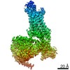 23281MC 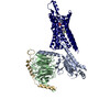 7ld3C M: map data used to model this data C: citing same article ( |
|---|---|
| Similar structure data |

-Links
 PDBj
PDBj


- Assembly
| Deposited unit | 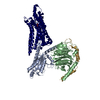
|
|---|---|
| 1 |
|
-Components
-Guanine nucleotide-binding protein ... , 3 types, 3 molecules ABG
| #1: Protein | Mass: 40502.863 Da / Num. of mol.: 1 Source method: isolated from a genetically manipulated source Source: (gene. exp.) Homo sapiens (human) / Gene: GNAI2, GNAI2B / Production host:  Trichoplusia ni (cabbage looper) / References: UniProt: P04899 Trichoplusia ni (cabbage looper) / References: UniProt: P04899 |
|---|---|
| #2: Protein | Mass: 38534.062 Da / Num. of mol.: 1 Source method: isolated from a genetically manipulated source Source: (gene. exp.) Homo sapiens (human) / Gene: GNB1 / Production host: Trichoplusia ni (cabbage looper) / References: UniProt: P62873 |
| #3: Protein | Mass: 7861.143 Da / Num. of mol.: 1 Source method: isolated from a genetically manipulated source Source: (gene. exp.) Homo sapiens (human) / Gene: GNG2 / Production host: Trichoplusia ni (cabbage looper) / References: UniProt: P59768 |
-Protein , 1 types, 1 molecules R
| #4: Protein | Mass: 43261.020 Da / Num. of mol.: 1 Source method: isolated from a genetically manipulated source Source: (gene. exp.) Homo sapiens (human) / Gene: CHRM4, ADORA1 / Production host: Trichoplusia ni (cabbage looper) / References: UniProt: P08173, UniProt: P30542 |
|---|
-Non-polymers , 2 types, 2 molecules 

| #5: Chemical | ChemComp-ADN /  Mass: 267.241 Da / Num. of mol.: 1 / Source method: obtained synthetically / Formula: C10H13N5O4 / Feature type: SUBJECT OF INVESTIGATION Mass: 267.241 Da / Num. of mol.: 1 / Source method: obtained synthetically / Formula: C10H13N5O4 / Feature type: SUBJECT OF INVESTIGATION |
|---|---|
| #6: Water | ChemComp-HOH / Mass: 18.015 Da / Num. of mol.: 1 / Source method: isolated from a natural source / Formula: H2O |
-Details
| Has ligand of interest | Y |
|---|---|
| Has protein modification | Y |
-Experimental details
-Experiment
| Experiment | Method: ELECTRON MICROSCOPY |
|---|---|
| EM experiment | Aggregation state: PARTICLE / 3D reconstruction method: single particle reconstruction |
- Sample preparation
| Component | Name: Human adenosine A1 receptor-Gi2-protein complex bound to its endogenous agonist adenosine Type: COMPLEX / Entity ID: #1-#4 / Source: RECOMBINANT |
|---|---|
| Source (natural) | Organism: Homo sapiens (human) |
| Source (recombinant) | Organism: Trichoplusia ni (cabbage looper) |
| Buffer solution | pH: 7.5 |
| Specimen | Embedding applied: NO / Shadowing applied: NO / Staining applied: NO / Vitrification applied: YES |
| Vitrification | Instrument: FEI VITROBOT MARK IV / Cryogen name: ETHANE-PROPANE / Humidity: 100 % / Chamber temperature: 277 K |
- Electron microscopy imaging
| Experimental equipment |  Model: Titan Krios / Image courtesy: FEI Company |
|---|---|
| Microscopy | Model: FEI TITAN KRIOS |
| Electron gun | Electron source:  FIELD EMISSION GUN / Accelerating voltage: 300 kV / Illumination mode: OTHER FIELD EMISSION GUN / Accelerating voltage: 300 kV / Illumination mode: OTHER |
| Electron lens | Mode: BRIGHT FIELD / Calibrated magnification: 47170 X / C2 aperture diameter: 50 µm |
| Specimen holder | Cryogen: NITROGEN / Specimen holder model: FEI TITAN KRIOS AUTOGRID HOLDER |
| Image recording | Average exposure time: 8 sec. / Electron dose: 50 e/Å2 / Detector mode: COUNTING / Film or detector model: GATAN K2 SUMMIT (4k x 4k) |
| EM imaging optics | Phase plate: VOLTA PHASE PLATE |
| Image scans | Width: 3838 / Height: 3710 / Movie frames/image: 50 |
- Processing
| CTF correction | Details: Phase plate CTF correction / Type: NONE |
|---|---|
| 3D reconstruction | Resolution: 3.3 Å / Resolution method: FSC 0.143 CUT-OFF / Num. of particles: 716000 / Symmetry type: POINT |
